We the Kings bassist and YouTube celebrity Charles Trippy recently uploaded to YouTube a video of his brain surgery. This is an amazingly powerful thing to watch and I recommend taking the time to do so if you have any interest in the brain and/or medicine that you watch it.
It’s not an easy viewing though, so I warn you.
I came across this video on reddit, and I took a shot at explaining the procedure and details over there. Most of what I’ve learned is from my research (e.g., A, B, C) in working with neurosurgeons, neurologists, and the incredible patients who undergo these surgeries.
The full text of that discussion is below.
OK I’m gonna take a whack at explaining what’s going on here for people who are interested. Note that I’m not a medical doctor, but I’m pretty sure most of what I’m about to say is correct to a first approximation. I’ll fix any errors as I or others spot them.
Why’s he awake? Why are they asking him to move his hands? Stuff like that.
Before I explain anything though, can I just say how damn amazing this is? Like, let’s all take a moment to step back and recognise that a person, who would have been much worse off otherwise, put his life in the hands of a few highly skilled people and some amazing technology and let them open his skull and remove something from his brain. I’ve seen this many, many times, but it’s good to just remember how amazing of a time we live in even though we don’t have flying cars or jet packs or whatever. Every day people’s lives are saved by medical technology like this.
Also, Mr Trippy’s a pretty amazing guy for volunteering to share this with the world. I don’t think people appreciate this stuff enough. Good on you man. I’d love to use this video when I teach.
On to the details. From this image here you can see that the tumour is in his right frontal lobe:
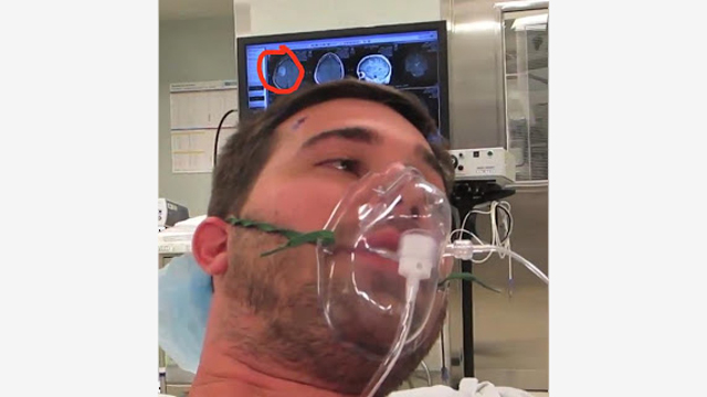
The image I’ve circled shows the tumour-related hyperintensity (bright whiteness) on a t2-weighted MRI, which allows you to differentiate tissues from fluids. In t2-weighted MRI, water is brighter. This particular kind of tumour makes the blood-brain barrier more permeable and therefore the region around it has more water and thus is brighter.
Note that I say the tumour is in the right half of his brain, but the tumour in the MRI image looks like it’s in the left. That’s because neurologists “flip” the MRIs so that left is right and vice versa. Supposedly it’s mirrored because that what the doctor sees when they’re looking at the patient.
Anyway, the fact that the tumour is in the back part of his right frontal lobe means that it’s close to the parts of the brain that control the muscles in the left half of his body. That is, the right motor cortex part of your brain innervates the muscles on the left side of your body and vice versa.
The reason the doctors keep saying stuff like “critical to move right now” and “how’s your hand doing there?” is because they want to minimise the possibility of cutting healthy brain tissue in his motor cortex. Neurosurgeons have a specific term for parts of the brain dealing with movement, speech, vision, and sensation. They call these regions “eloquent cortex”, and they’re very worried about damaging them… otherwise people could end up paralysed, blind, or unable to speak or understand speech.
So they’re really watching him very carefully, and that’s why he’s awake. They’re making sure he’s moving his hand just fine.
Given the location of the tumour, prior to his surgery they probably did what’s called a “wada test” to see if his language functions are localised to the left or right hemisphere of his brain. If he’s right handed, as a male he’s got a 99% or so probability of language functions being in his left hemisphere, but usually surgeons check to make absolutely sure (right-handed women are about 95% left-hemisphere language dominant). Functional brain imaging (EEG, fMRI, MEG) just isn’t quite good enough on a single person for surgeons to really be 100% positive. Whereas the effects of the wada test are… well… pretty conclusive.
The wada test is essentially using a barbiturate injected directly into the brain via the carotid artery to put half the brain to sleep. If they inject the barbiturate into the left hemisphere of your brain, and you’re left hemisphere language dominant, then… well you’ll feel funky for a while and have some language issues.
I’ve only half-jokingly asked my neurosurg friends to do this to me just to see what it feels like. Apparently “do no harm” and ethics and stuff, so no go so far.
Oh! Right at the beginning of the video a woman says “you’ll feel a pinch and burn”. I’m pretty sure she’s doing a local anaesthetic to the skin to numb it so they can screw the stereotactic frame into his skull. This lets them keep all of their instruments correctly positioned and still even though he’s awake, talking, and moving around a little.
That’s all I can think of for now. If anyone notices anything that I didn’t address and they’re curious about, ask me. If I don’t know, I’ll bug my neurosurg friends and update this post with answers.
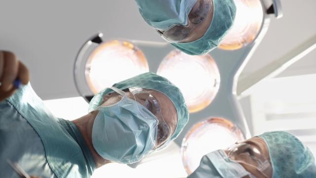
Picture: Shutterstock/bikeriderlondon
This post was originally published on Oscillatory Thoughts, a neuroscience blog written by Bradley Voytek, PhD, Professor of Cognitive Science and Neurosciences, UC San Diego. You can follow Dr Voytek on Twitter here.
This post has been republished with permission.