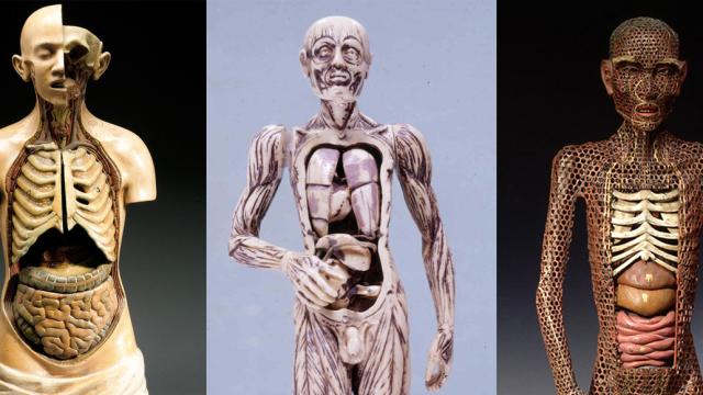
Since the early days of medicine, it’s been a huge challenge to communicate anatomical discovery with an audience without any actual human dissection. But thanks to the creativity of the medical experts and artists, a huge variety of anatomical models: From wax figures and ivory manikins to papier mache dolls and multi-colour portfolio prints.
The following beautiful and morbid pieces were made from the highest scientific and artistic design, and range from small organs to full anatomical figures, from the Middle Ages to the 20th century. They often consist of removable parts that could be “dissected” to reveal the inner workings of the mysterious human body.
Viewer discretion is advised below this point.
This is an extremely rare Japanese model of a human body made of copper, lacquer, wood and other materials, from 1669. The model combines Asian and Western medical knowledge.
Picture: Museum of Ethnology, Hamburg/Virtual Collection Of Asian Masterpieces
An advertising brochure by Bayer, c1930, overlays the bodily systems on each page.
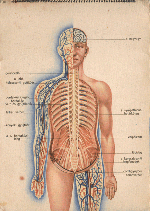
Picture: Attila Nagy/Gizmodo
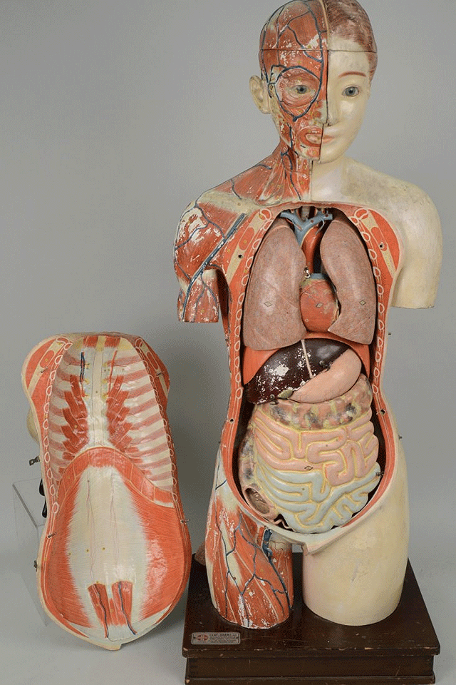
A 72 inch-tall polychrome plaster anatomical model has removable arms, and the torso opens to reveal removable organs. It was made in the early 20th century by Maison Deyrolle in Paris.
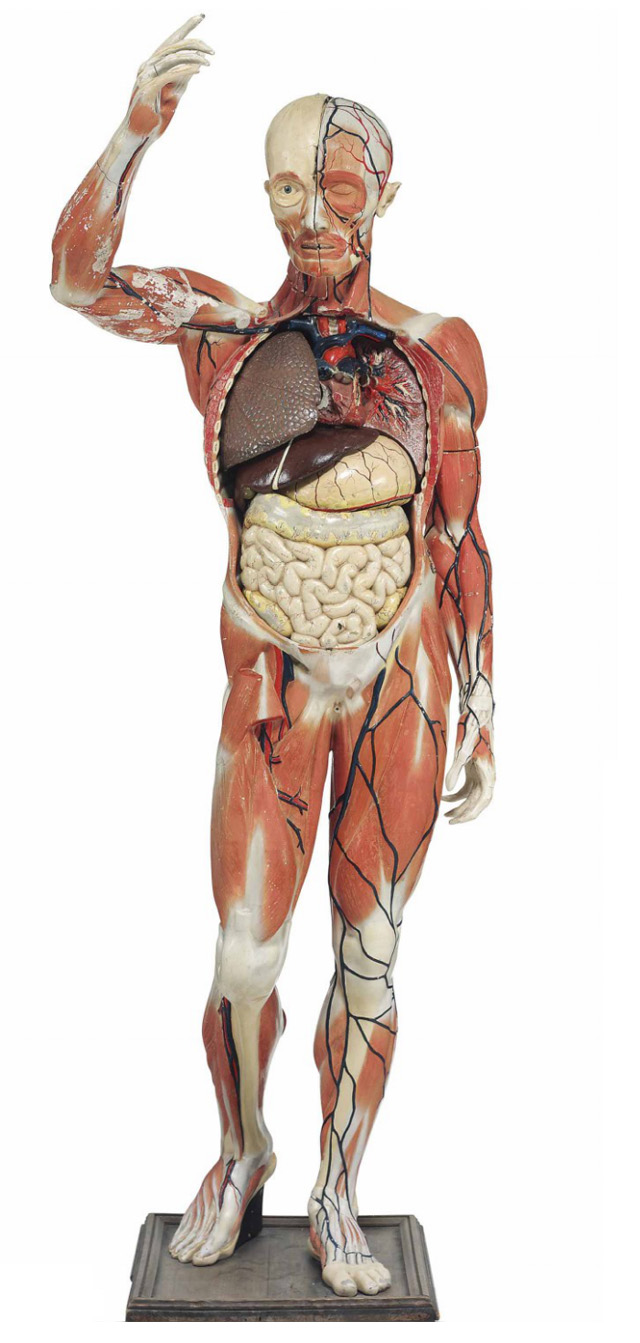
Picture: Christie’s
A carved ivory, wood and glass anatomical model of an eye. German, probably late 17th century.
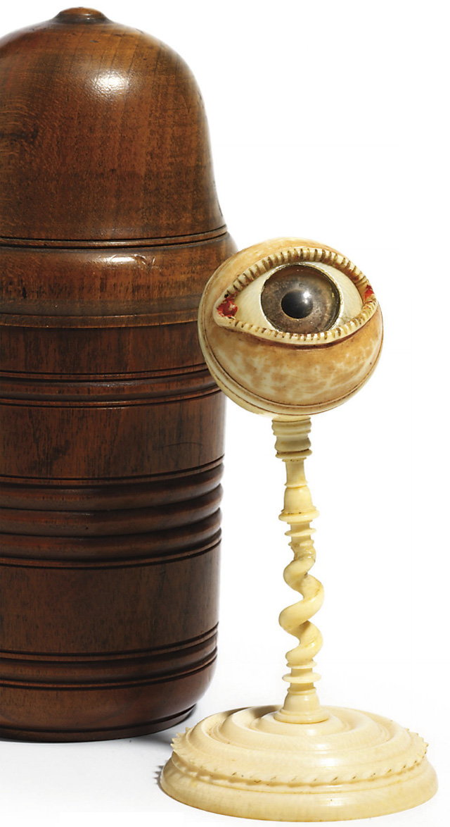
Picture: Christie’s
A carved boxwood model of a head and the facial muscles, from 19th century Italy, sits only three inches tall.
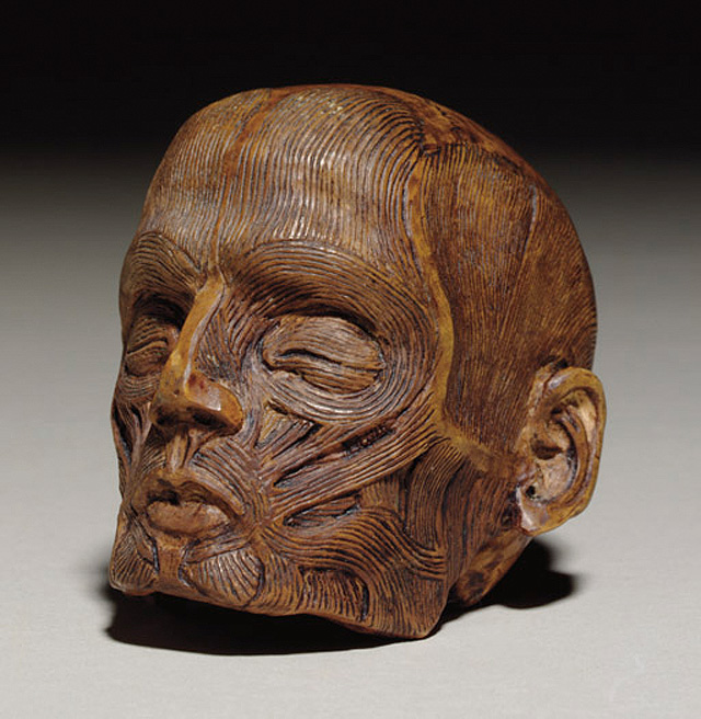
Picture: Christie’s
A life-sized, intricately-detailed papier-mâché anatomical model, devised and created by the French physician Dr. Louis Thomas Jerome Auzoux, circa 1882.
Picture: Bonhams
A wax anatomical half model of the torso of a pregnant woman, from early 19th century France.
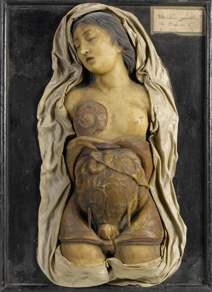
Picture: Bonhams
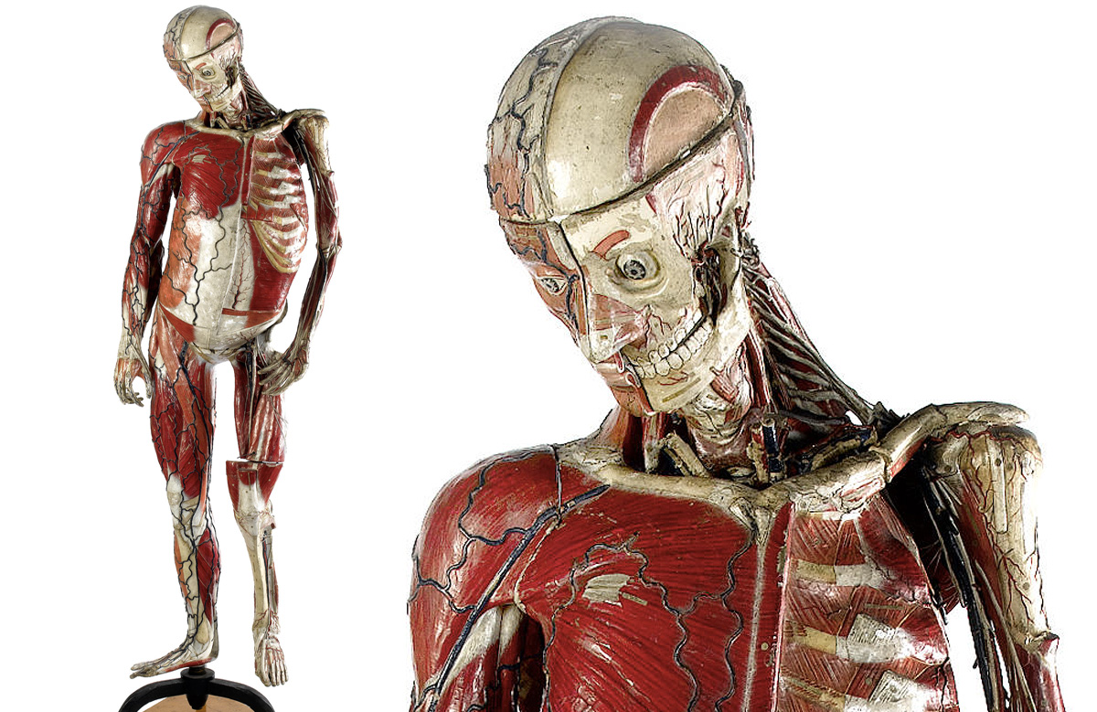
A three quarter length male anatomical model, probably French, 19th century.
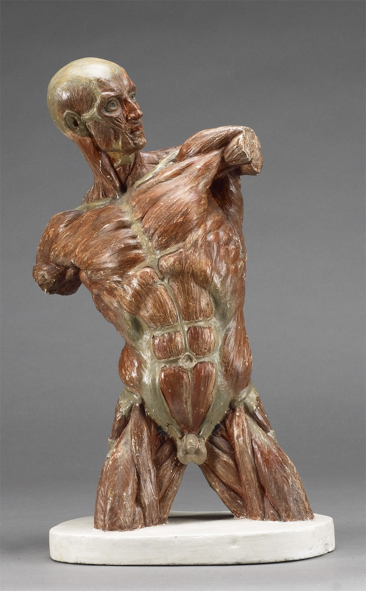
Picture: Bonhams
An Adam Rouilly & Co. painted plaster anatomical model of a male torso. English, circa 1900.
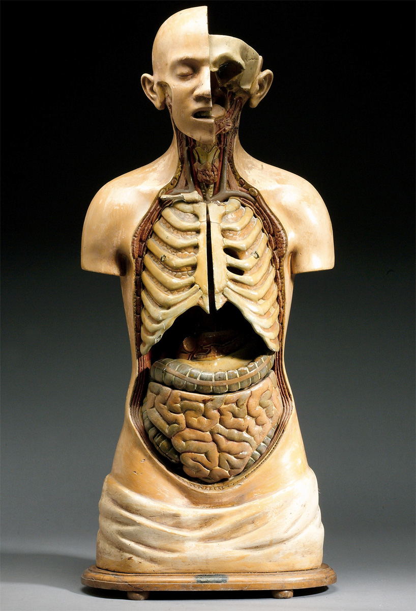
Picture: Bonhams
Anatomical models of a male body with chromolothograph cut-outs and overlay displays, edited by W.S.Furneaux, published by George Philip & Son, London.
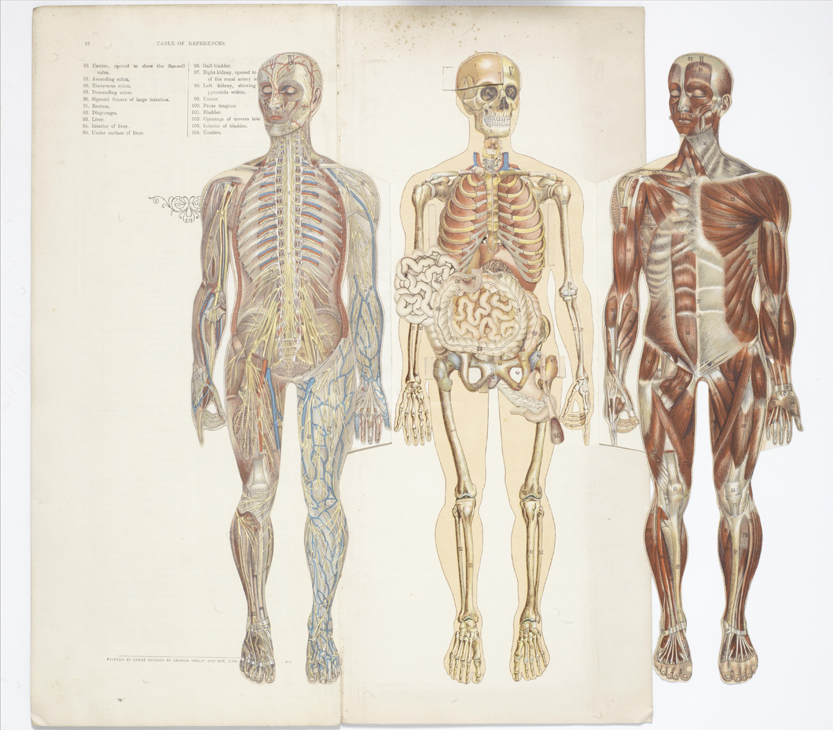
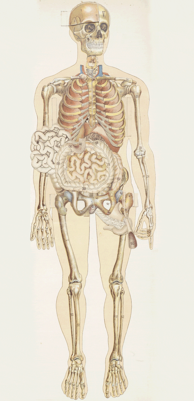
Picture: Bonhams
This anatomical rubber and bone model of an infant skeleton dates from the 1920s or ’30s.

Picture: Bonhams
An antique carved Chinese human acupuncture model.
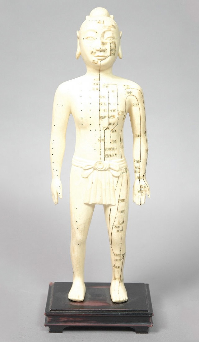
Picture: Live Auctioneers
Smith’s outlined paper maps of the human system, published by American Manikin Co., Peoria, Illinois, 1888.

Picture: Live Auctioneers
Anatomical figure of a woman, made by Clay-Adams Co., Inc., New York.
Picture: Live Auctioneers
Life-size chromolithographed manikin with numerous overlays revealing the anatomical make-up of a man, mounted on board frame. Publisher: James T. White, New York, 1886.
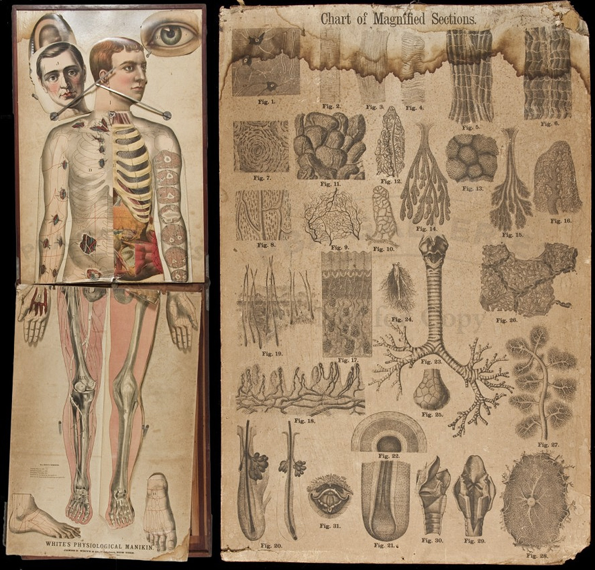
Picture: Live Auctioneers
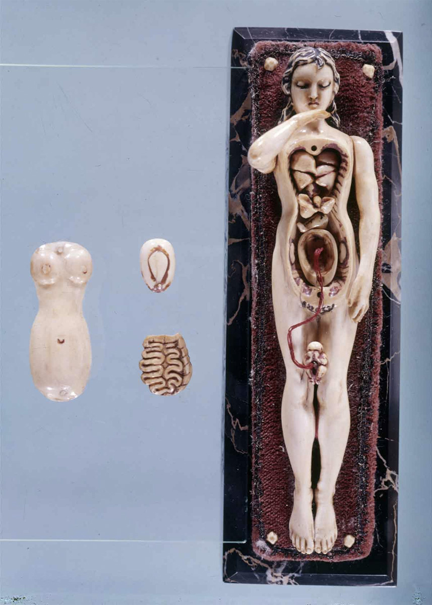
These male and female anatomical dolls, made of ivory, are each less than seven inches tall. Circa 1400.
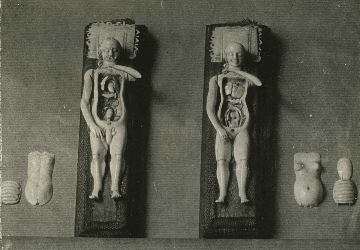
Picture: Otis Historical Archives National Museum of Health and Medicine
A grotesque anatomical and astrological doll at the Museum of Hygiene, Munich, Germany.
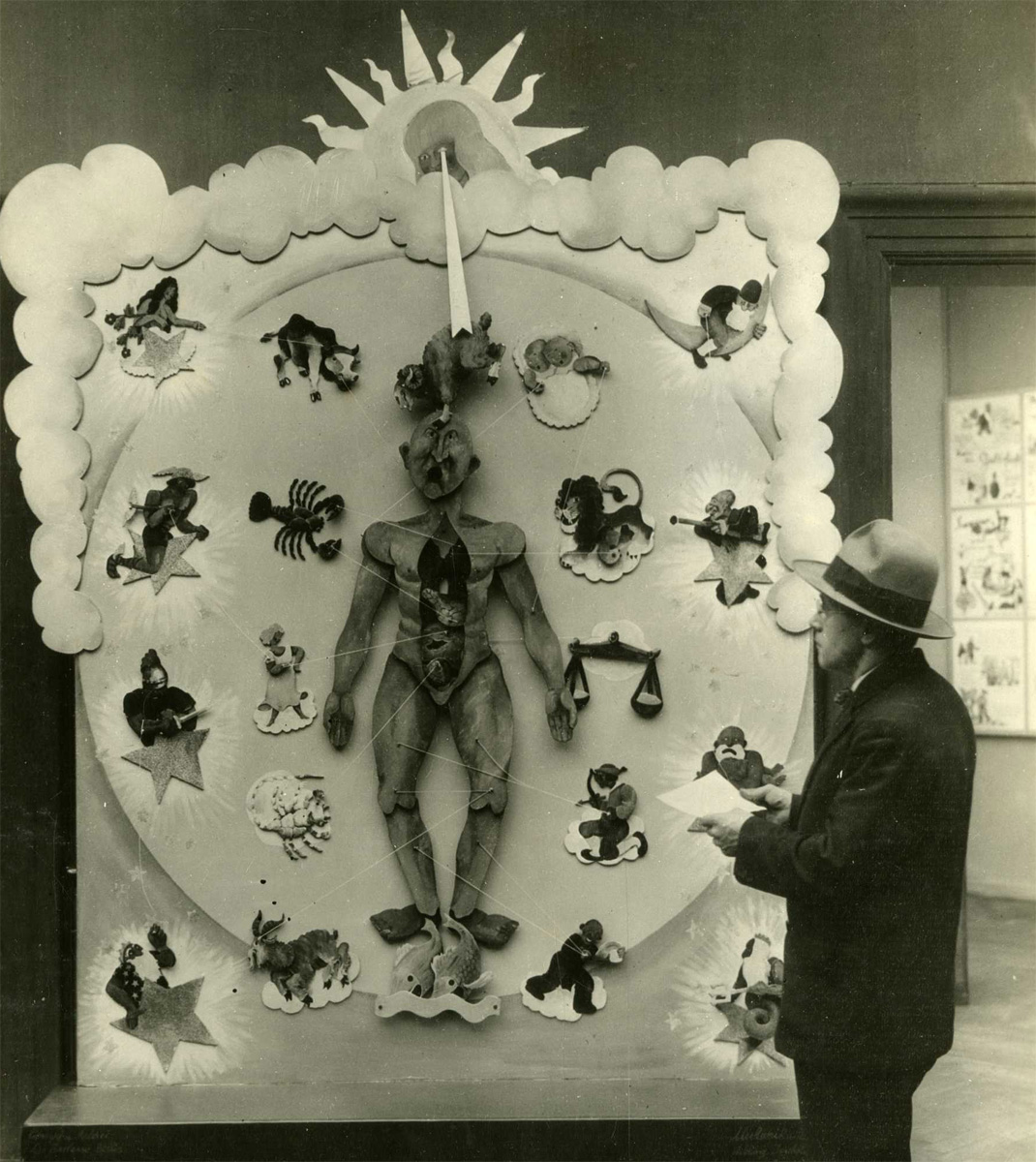
Picture: Otis Historical Archives National Museum of Health and Medicine
Ivory anatomical model of a pregnant woman at the Deutsche Historishes Museum.
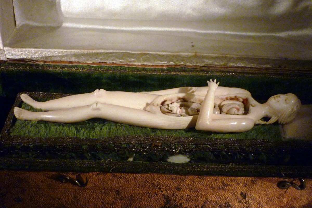
Picture: Will Manley
Dr. Auzoux’s papier-mâché pregnancy model at the Museum Boerhaave, Leiden, the Netherlands. Circa 1875-1900.
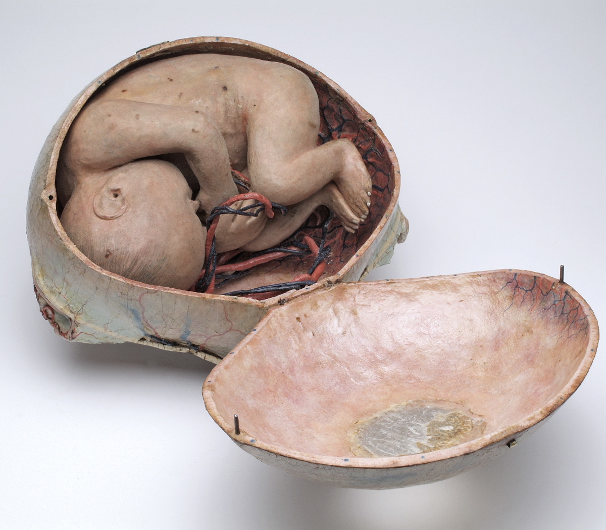
Picture: Museum Boerhaave/Wikimedia Commons
A mid-19th century wax specimen by Joseph Towne, at the Gordon Museum, Kings College, London, UK.
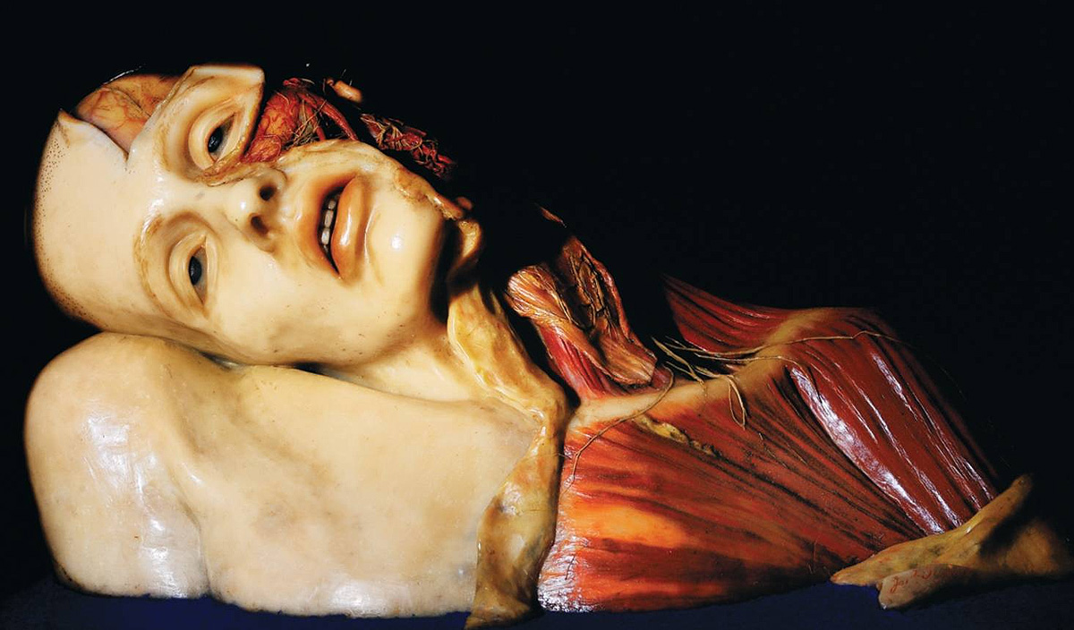
Picture: Anatomical Society of Great Britain and Ireland
“Anatomical Venuses” were life-sized wax anatomical models of idealised women, extremely realistic in appearance and often adorned with real hair and ornamental jewelry. This is “La Venerina,” a wax anatomical model by Clemente Susini, from the 18th century.
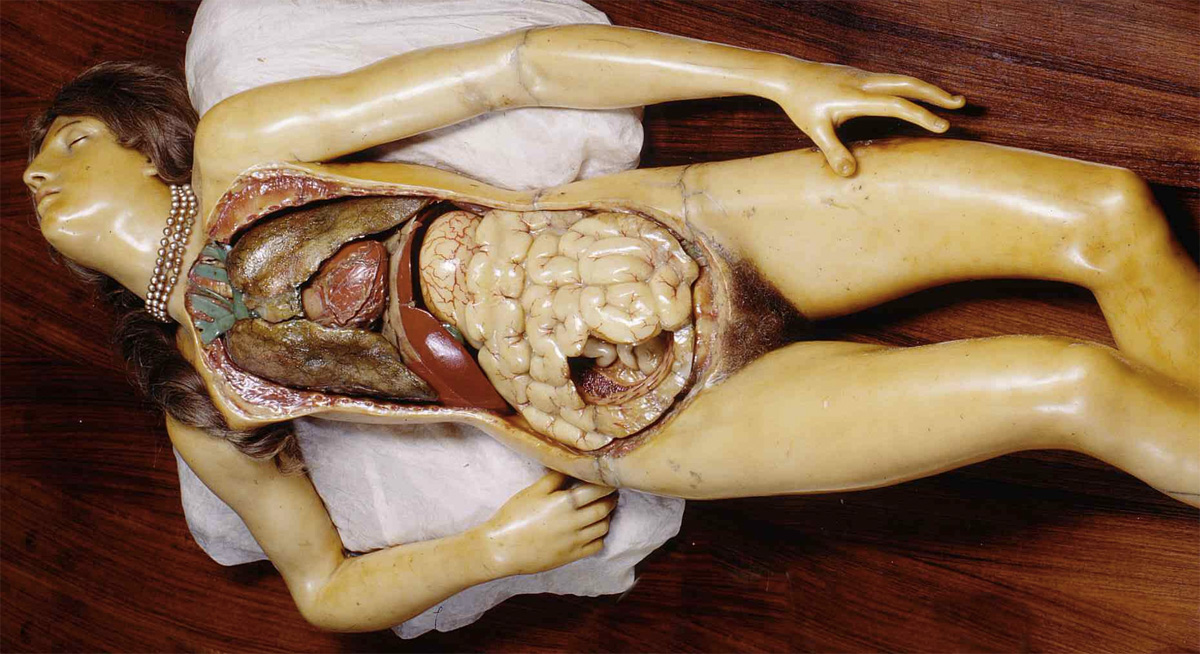
Picture: Museo delle Cere anatomiche “Luigi Cattaneo”
These 19th century pregnant dolls from Japan were created primarily to teach midwives how to deliver babies, but they were probably used for entertainment purposes as well.
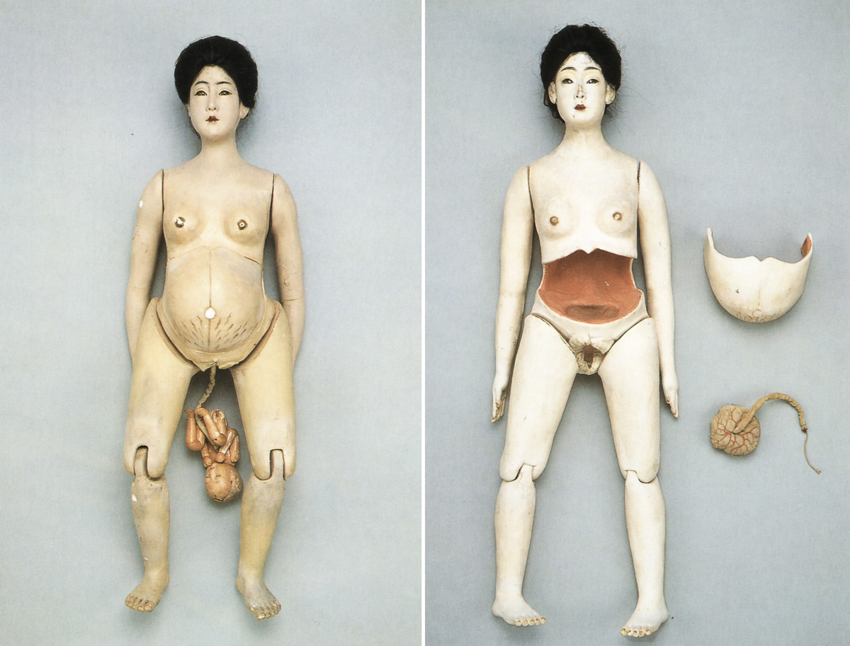
Picture: Geijutsu Shincho magazine, July 2001/Pink Tentacle
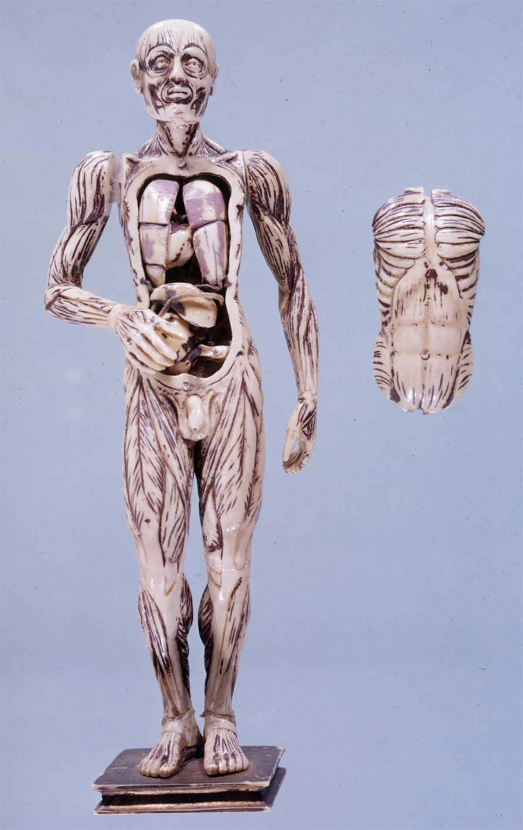
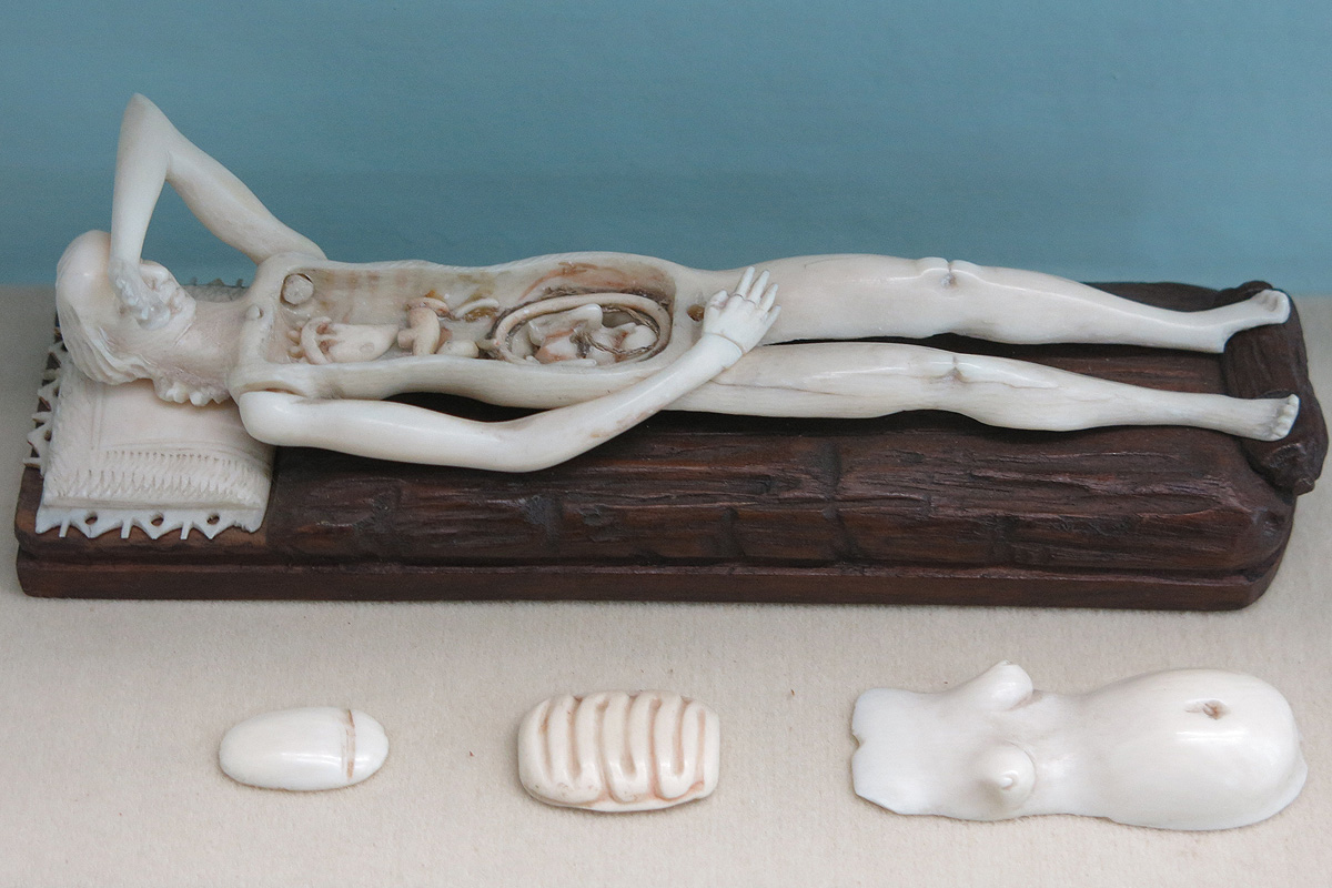
These manikins, between 6 to 7 inches in length, were made from solid pieces of ivory in 16th through 18th century Europe. The arms were carved separately and are moveable. The thoracic and abdominal walls can be removed, revealing the viscera. In some manikins, the internal organs are carved in the original block and are not removable, while here they are formed into separate pieces that can be removed.
Stephan Zick’s anatomical manikin made of ivory, mid-17th century. From the collection of the Semmelweis Museum, Budapest, Hungary.
Picture: Attila Nagy/Gizmodo
Male anatomic torso with exposed ribs and muscles. Carved Linden wood, early 18th century. From the collection of the Semmelweis Museum, Budapest, Hungary.
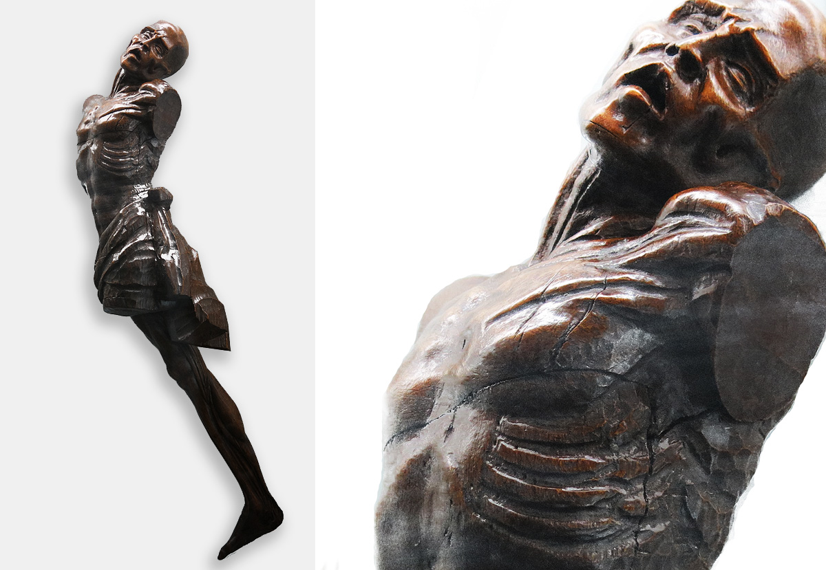
Picture: Attila Nagy/Gizmodo
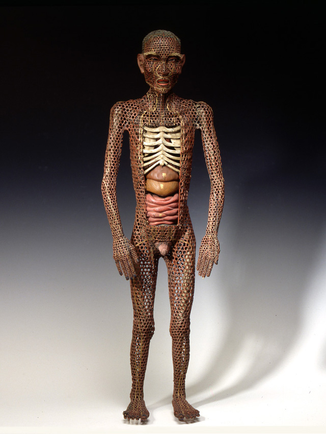
Demonstration doll for the presentation of the acupuncture points (China, 20th century). From the collection of the Semmelweis Museum, Budapest.
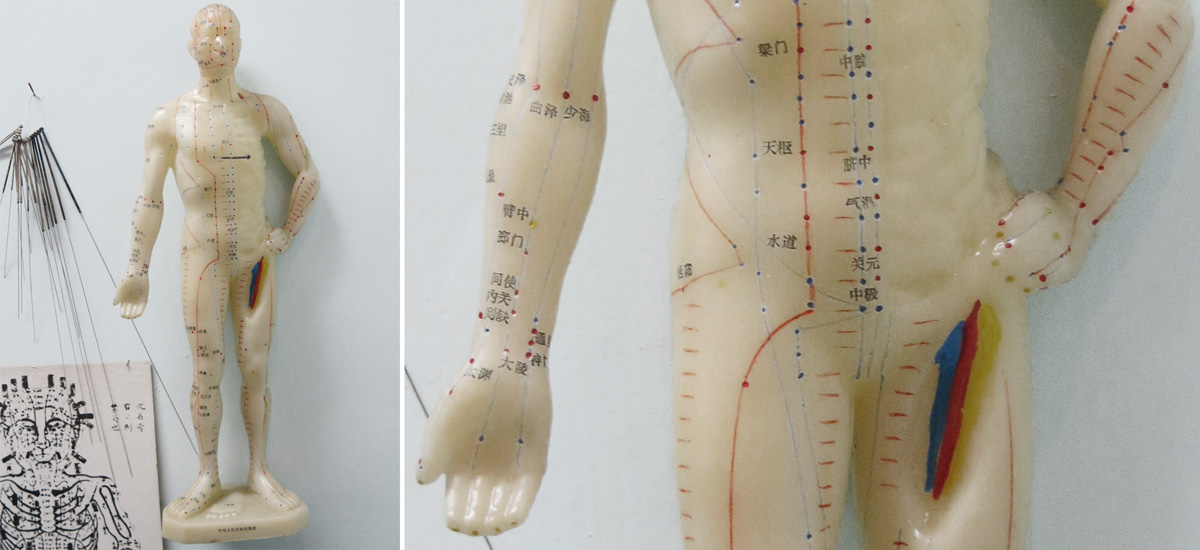
Picture: Attila Nagy/Gizmodo
Here’s another life-sized wax “anatomical Venus,” from the workshop of Felice Fontana, Florence, Italy, circa 1780-1785. From the collection of the Semmelweis Museum, Budapest.
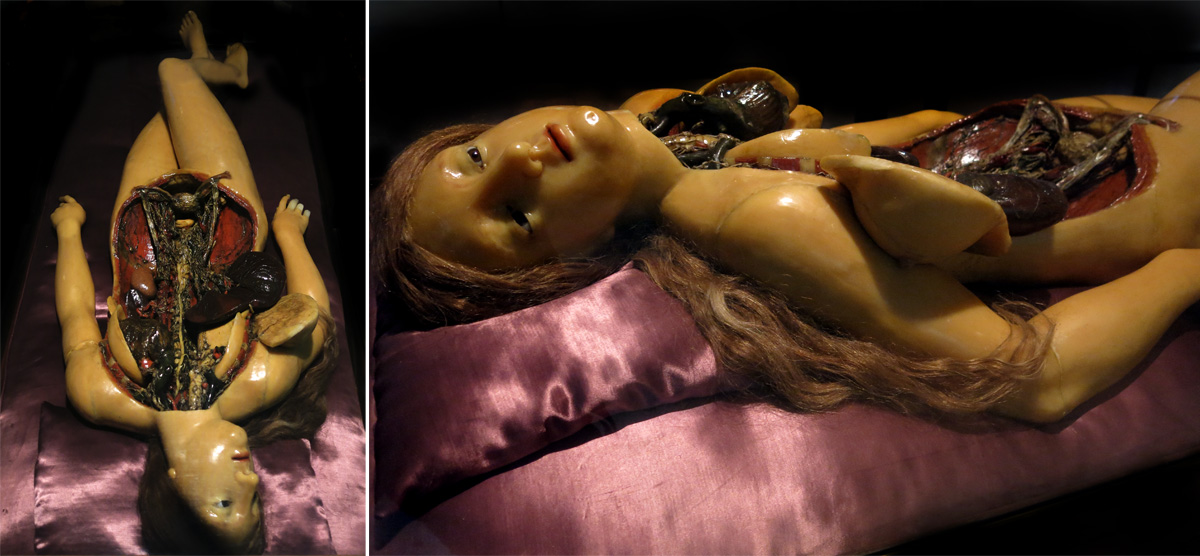
Picture: Attila Nagy/Gizmodo