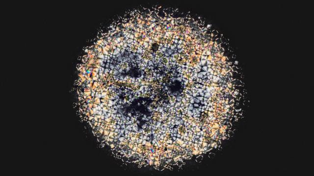
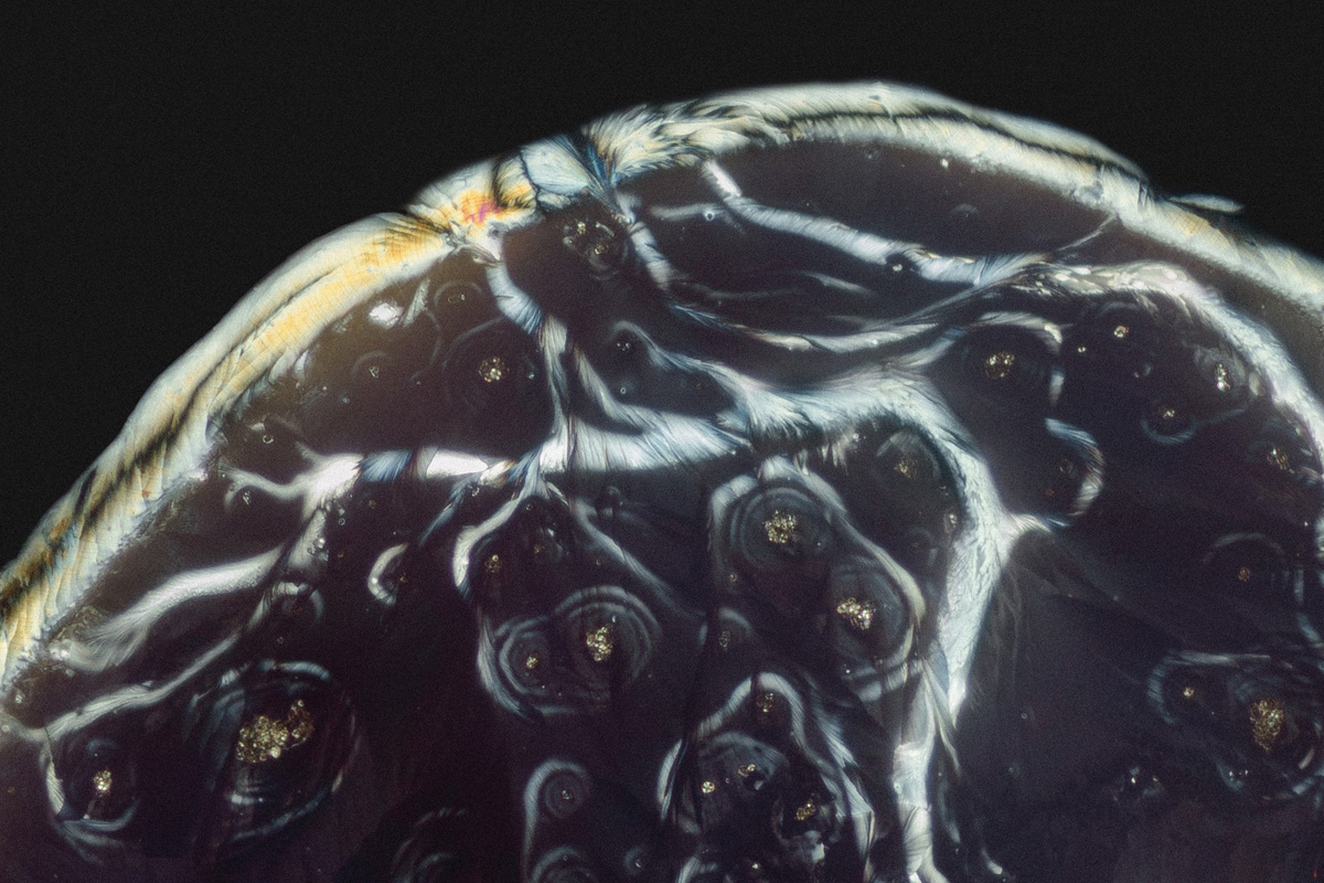
These stunning macro photos — micrographs — captured by Dutch photographer Maurice Mikkers are going to show you a whole new world, which still seems familiar on many levels.
I have to admit, I have never thought about how tablets and pills look like if we take samples of them and put them under microscope. But Maurice Mikkers — a licensed Medical Laboratory Analyst by the way — had a lot of unanswered questions and wanted to see and capture the crystal structures of pill agents by himself. And the results of his curiosity are truly amazing.
You can read about the whole work process in this detailed Vantage article:
Each prospective sample is crushed to powder in mortar and pestle and weighed out in uniform measurements. The powder is increasingly diluted to afford a wide selection of material to shoot. Applying heat speeds up the evaporation process and seems to yield more aesthetically appealing crystals, but slides still air dry under Mikkers’ scrutiny. Favoured results are duplicated to create an experiment group.
After calibrating both the camera and the microscope Mikkers fires off frames at different ISO and speeds, adjusting the angle of the slide for different perspectives on the crystalline structures. Once satisfied, he shoots in HDR a comprehensive grid of the entire sample. The images are stiched together later in digital post production.
Throughout, the entire workflow, Mikkers adheres to something akin to rigorous scientific method — samples are tracked in Excel. Meticulous notes are kept. Control groups are maintained. Like all good science, he wants to be able to repeat the process and, hopefully, the results.
What amazes me is that I feel I have seen the following images many times. Stellar clouds, galaxies, diatoms, snowflakes, aerial photos of our planet and crystal LSD? All the same.
Potassium bitartrate
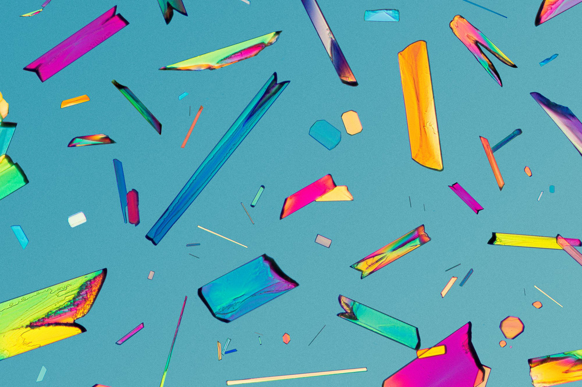
Diclofenac
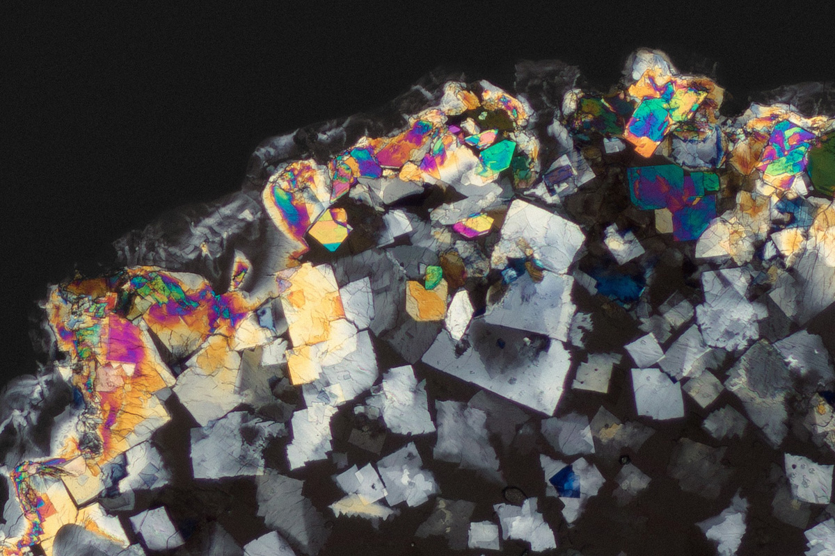
Amphetamine
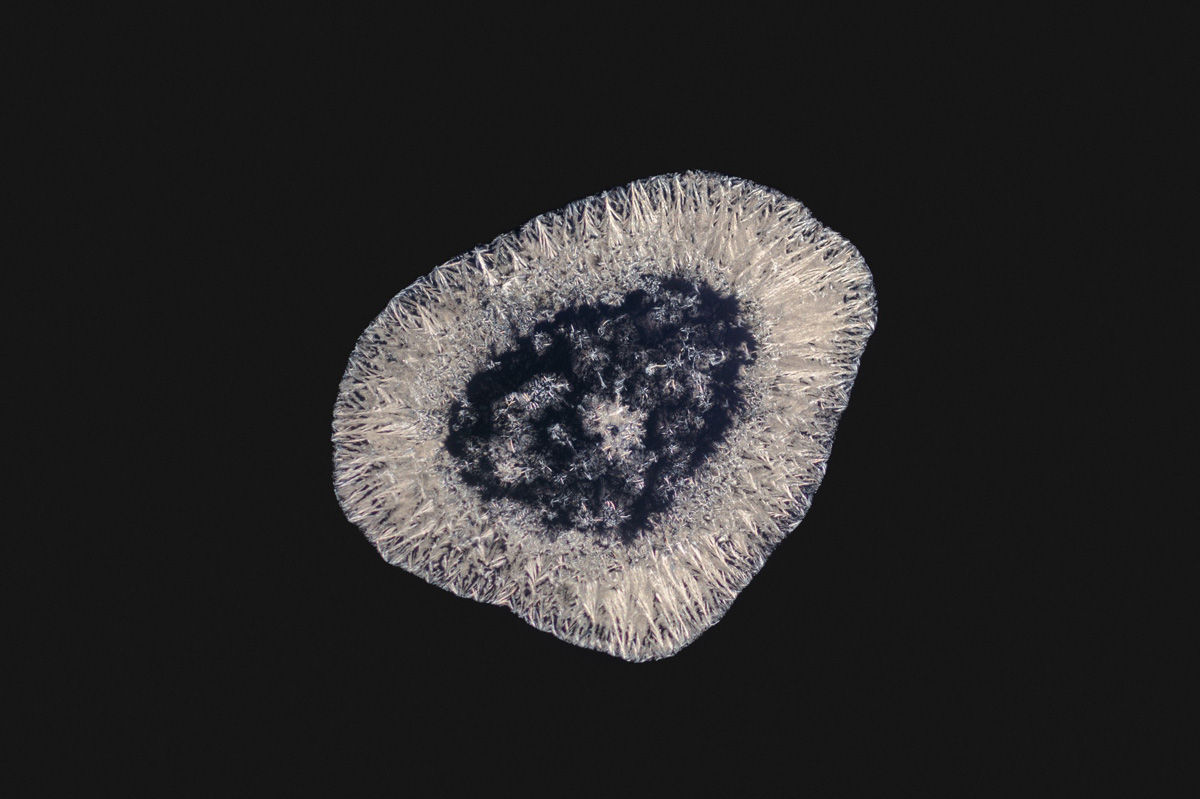
2C-B
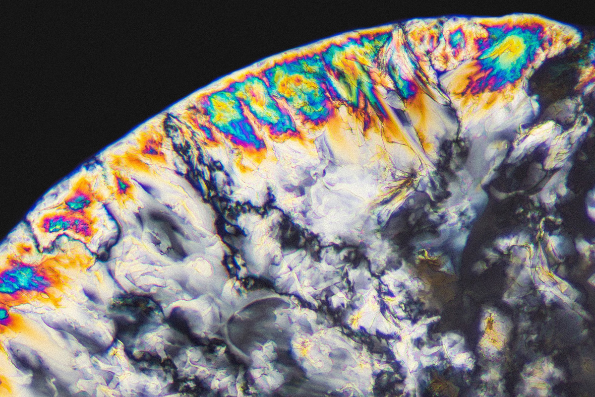
Diltiazem
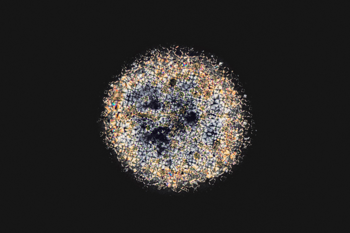
Birth control pill
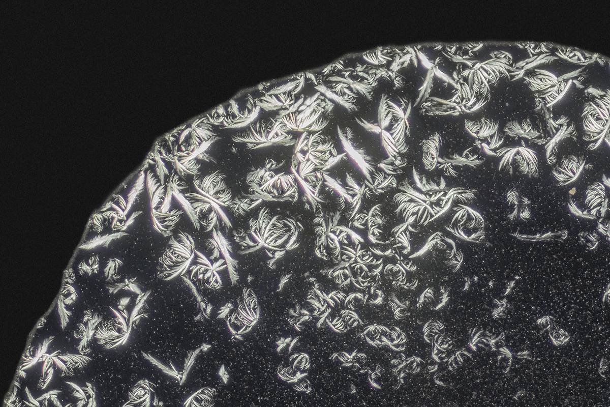
Gamma-Hydroxybutyric acid
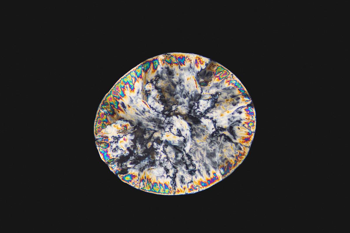
Vitamin C
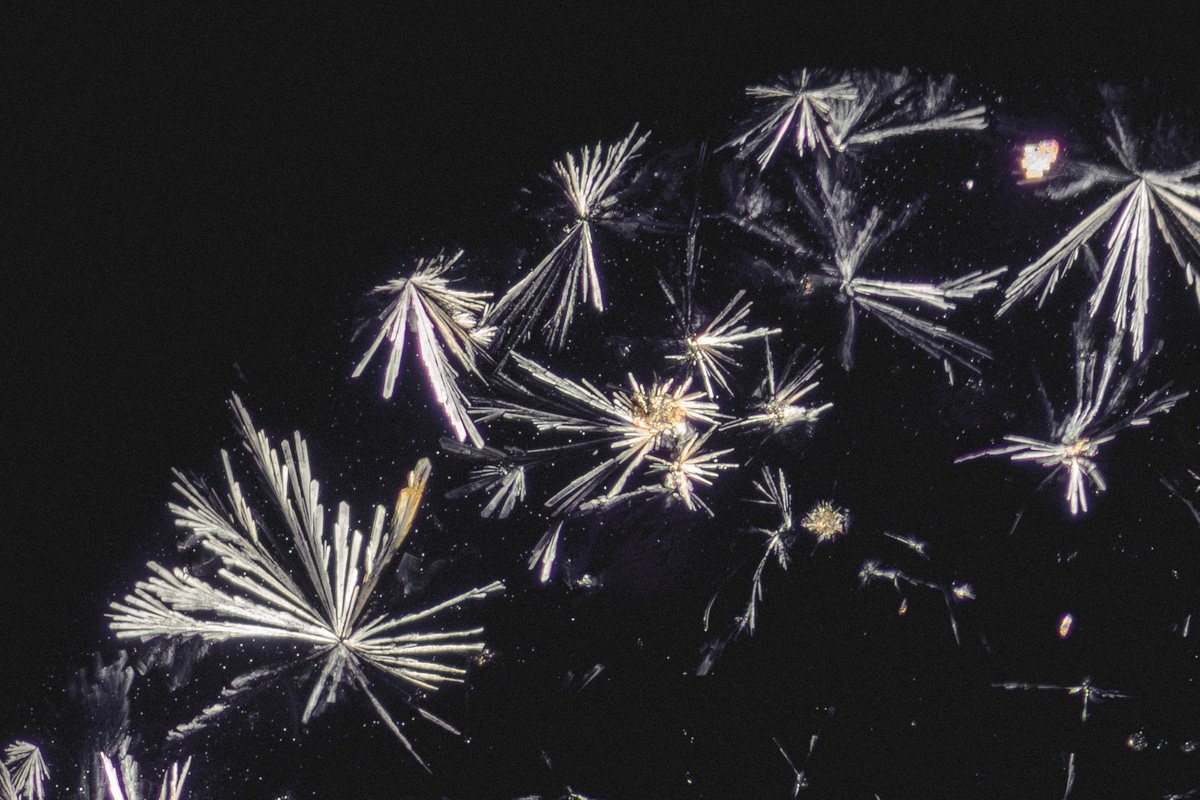
Ciprofloxacin
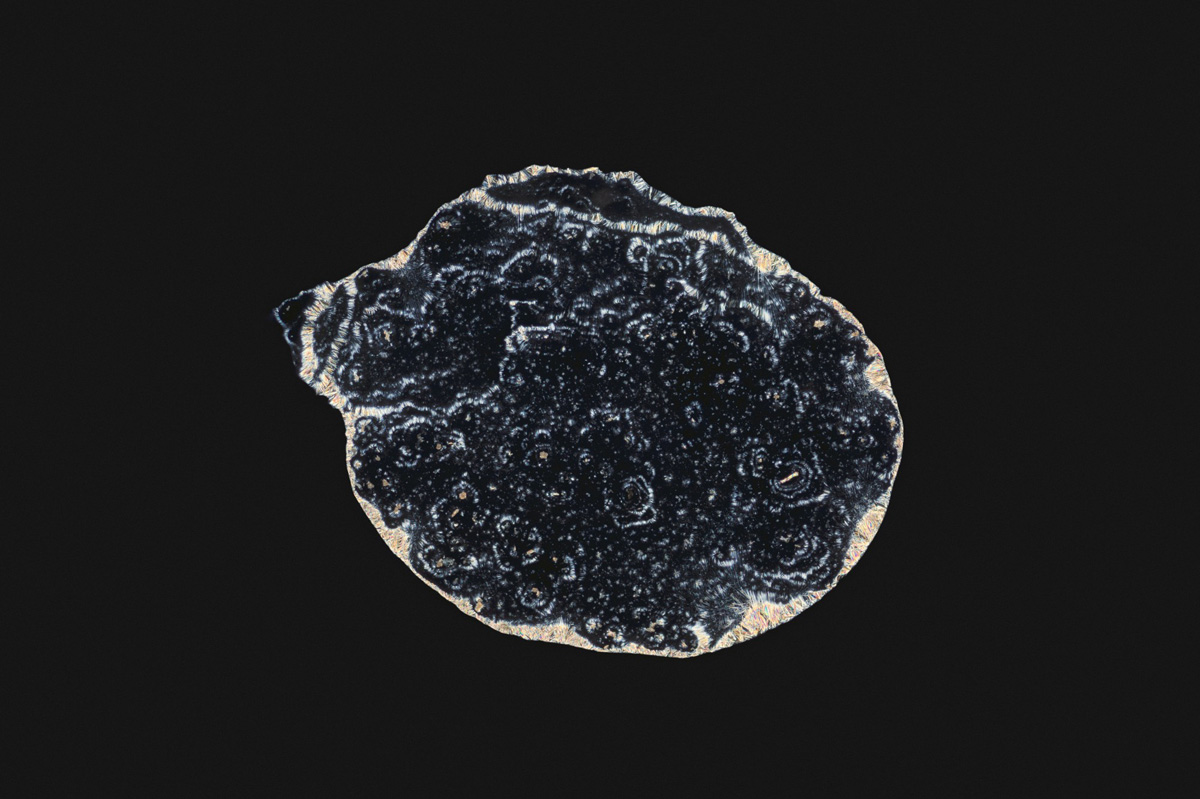
N,N-Dimethyltryptamine
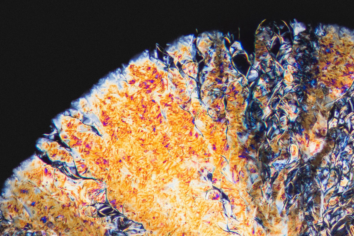
Dipyridamole
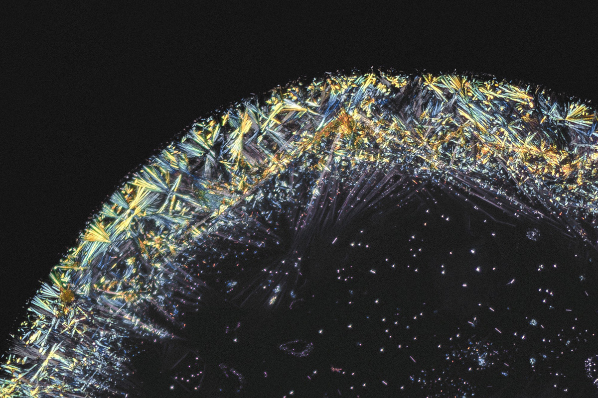
Lysergic acid diethylamide
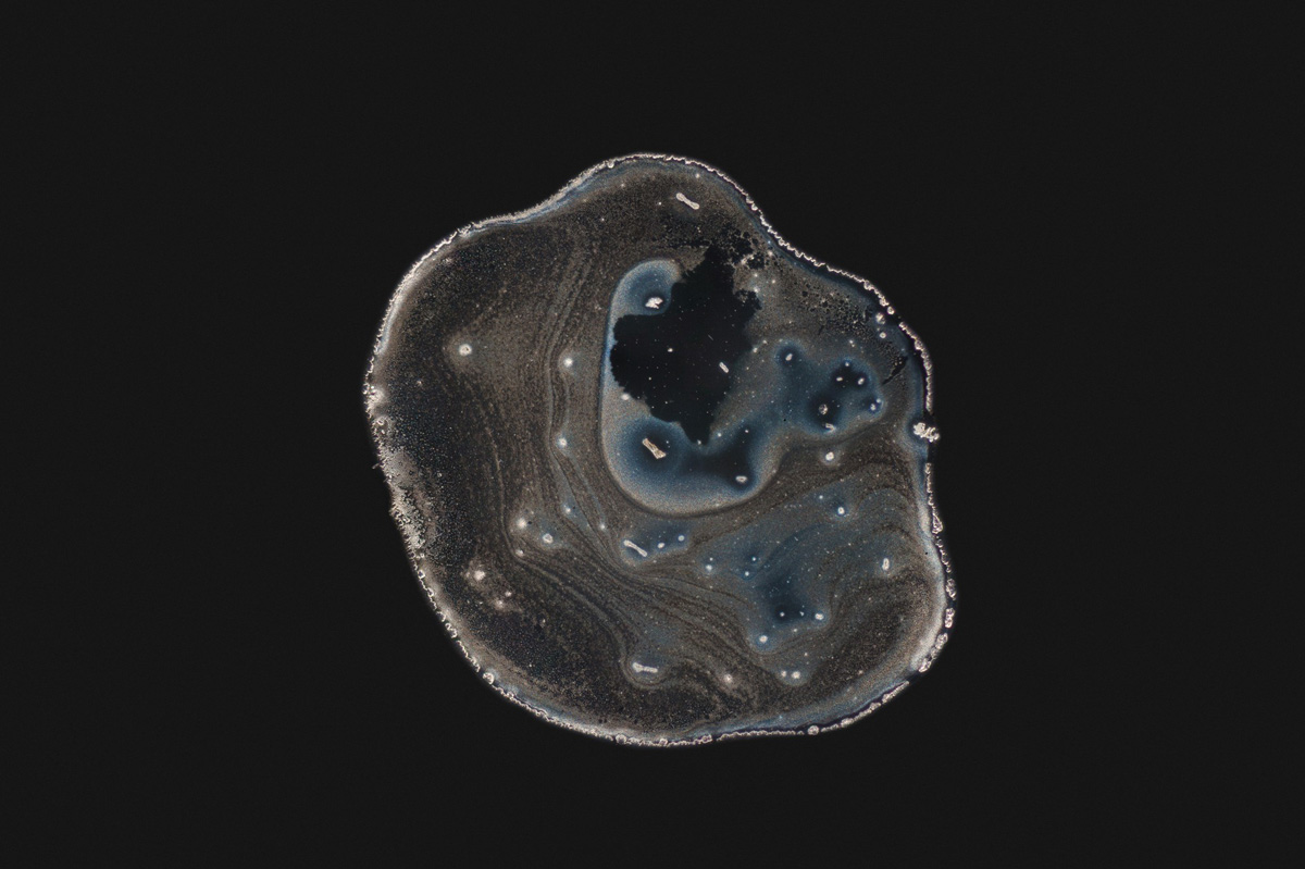
Acetaminophen
http://www.twitter.com/mauricemikkers
