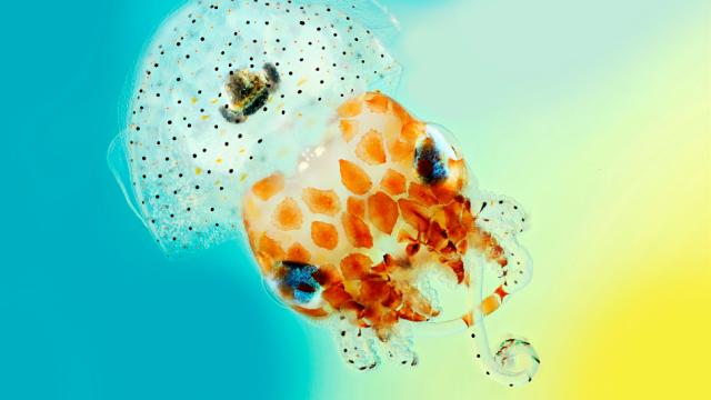
The finalists of the 2017 Wellcome Image Awards have been announced, showcasing the best science-related imagery from the past year. This year’s crop features a bioluminescent squid, a high-tech contact lens and a microscopic “brain” on a chip.
A bioluminescent Hawaiian bobtail squid. (Credit: Mark R Smith, Macroscopic Solutions)
The winners will be announced on March 15 at the Wellcome Trust in London. Judges will be evaluating the images for quality, technique, visual impact and their ability to communicate and engage. Here are our favourites.
Vessels of a Pig Eye
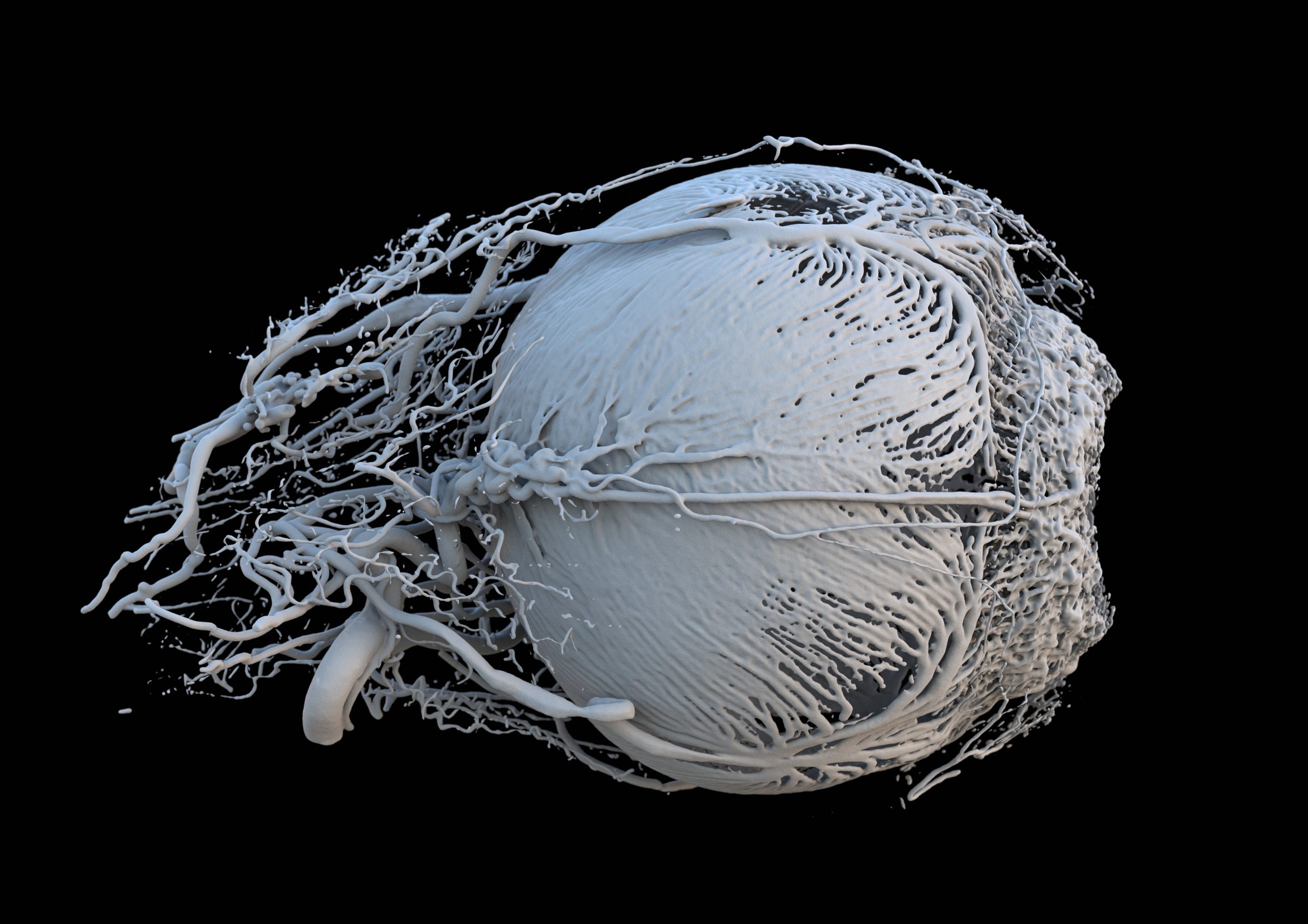
Credit: Peter M Maloca, OCTlab at the University of Basel and Moorfields Eye Hospital, London; Christian Schwaller; Ruslan Hlushchuk, University of Bern; Sébastien Barré
Using computed tomography (CT) and 3D printing, researchers from Switzerland created this unique look at blood vessels within a pig’s eye. These vessels bring energy and food to the muscles surrounding the iris, which controls how much light enters the eye. The pupil is located on the far right.
Language Pathways of the Brain
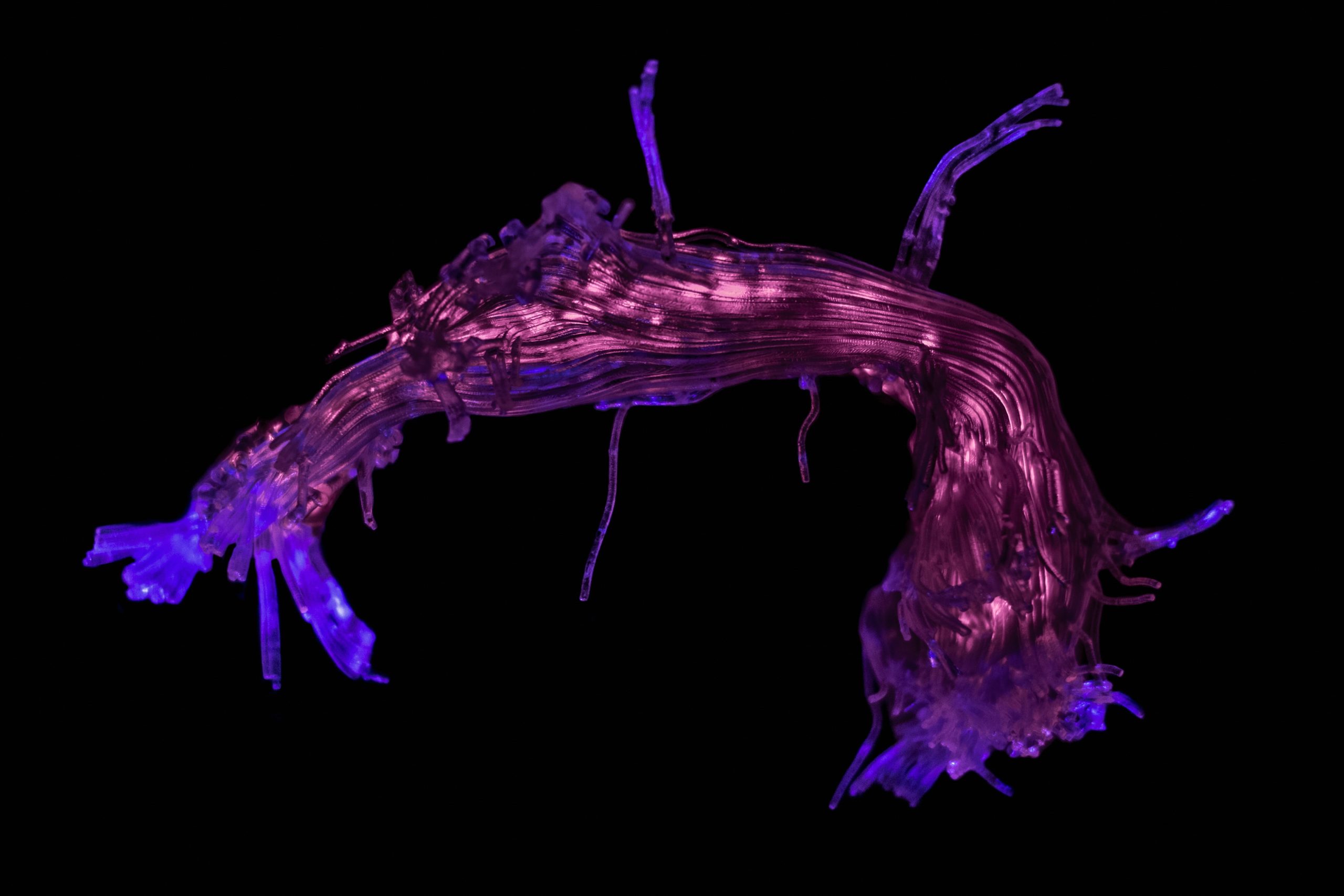
Credit: Stephanie J Forkel and Ahmad Beyh, Natbrainlab, King’s College London; Alfonso de Lara Rubio, King’s College London
A 3D-printed reconstruction of the white matter pathway connecting areas for speech and language. This model was created with a technique called tractography, which uses a scanner to track the movement of water molecules within the brain.
Surface of a Mouse Retina
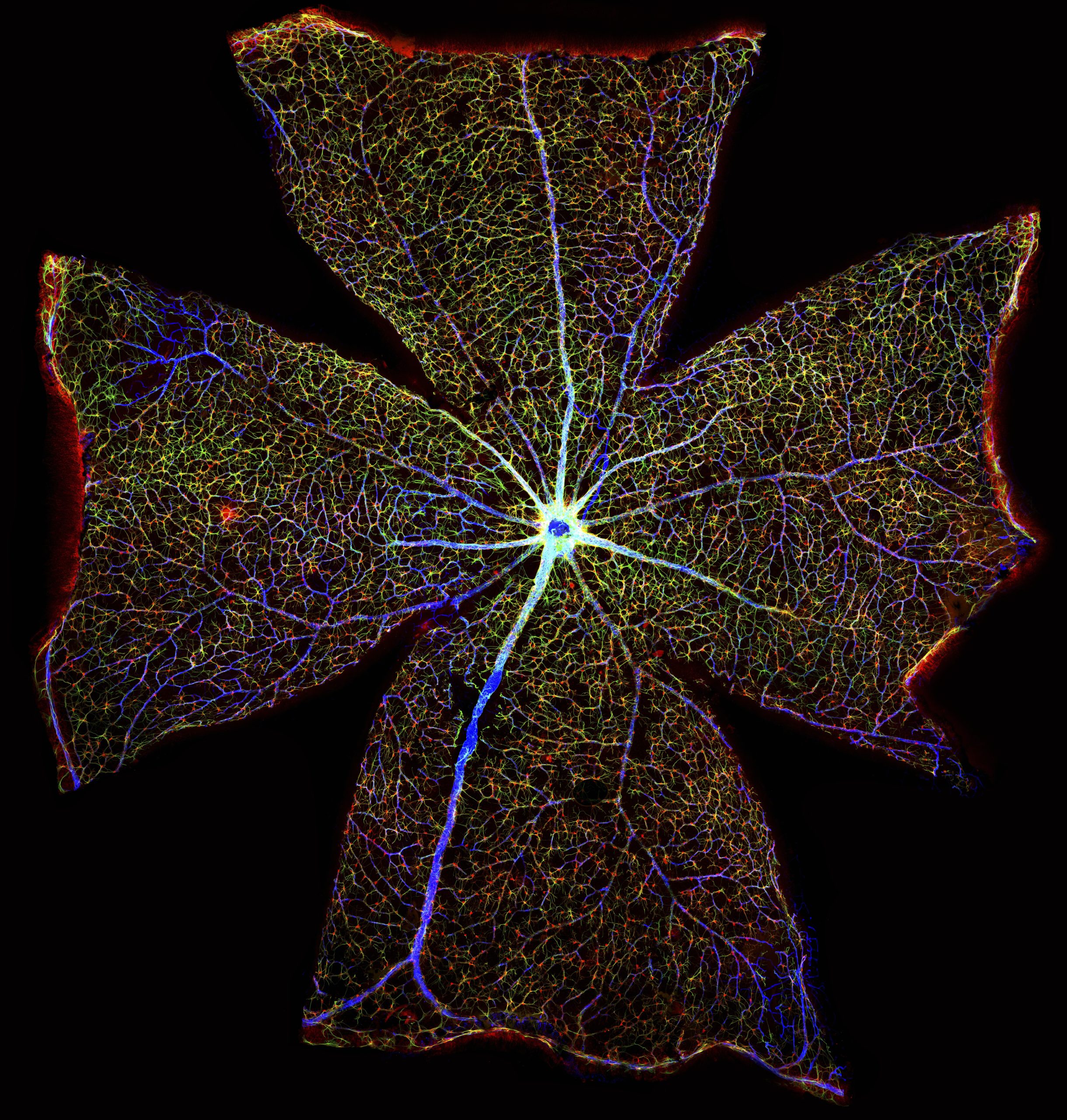
Credit: Gabriel Luna, Neuroscience Research Institute, University of California, Santa Barbara
Over 400 microscopic images were stitched together to create this view of a mouse’s retina. The lines in blue are blood vessels, and astrocytes (specialist cells of the nervous system) are shown in red and green. By researching the changing function of astrocytes during retinal degeneration, scientists are hoping to develop new treatments for vision loss.
The Placenta Rainbow
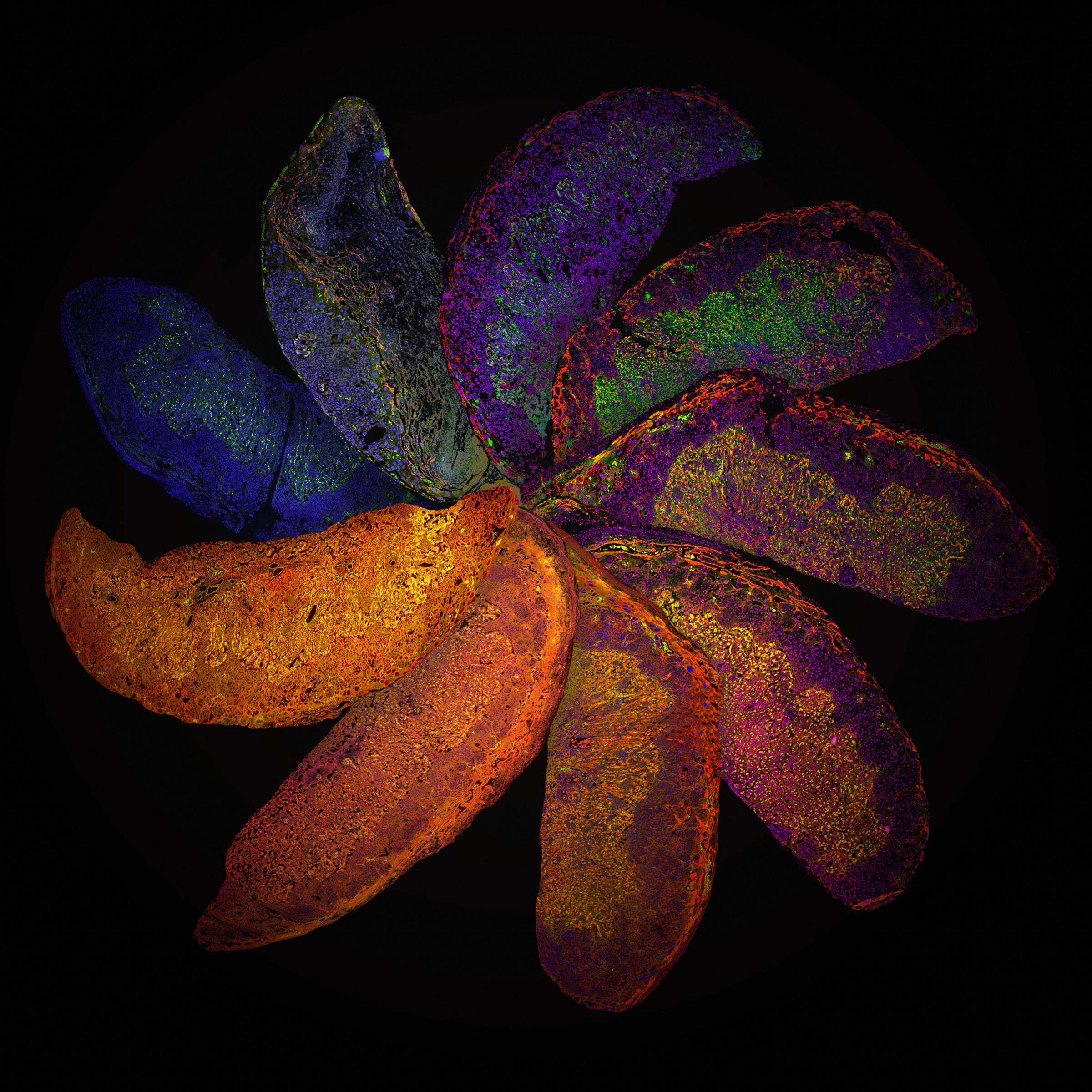
Credit: Suchita Nadkarni, William Harvey Research Institute, Queen Mary University of London
These placentas are from genetically-modified mice, each with their own distinct immune system. The resulting “Placenta Rainbow” highlights the differences in placental development that can result from the manipulation of the mother’s immune system, allowing researchers to better understand and treat complications that arise during human pregnancies.
Unravelled DNA in a Human Lung Cell
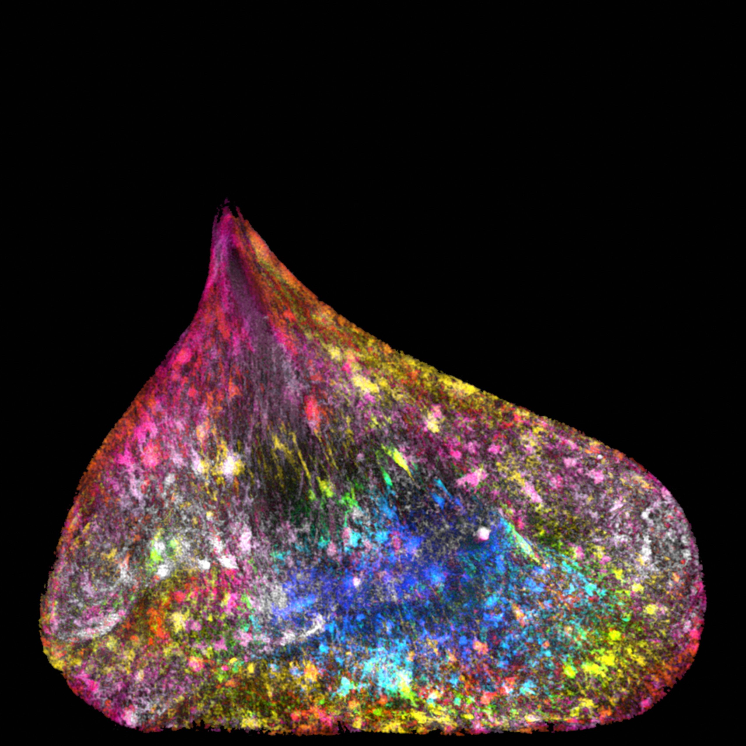
Credit: Ezequiel Miron, University of Oxford
The nucleus of a new human lung cell, packed with DNA. The cell’s deformed shape is the result of lingering tension, caused by rope-like strands of DNA being pulled between the two cells.
Developing Spinal Cord
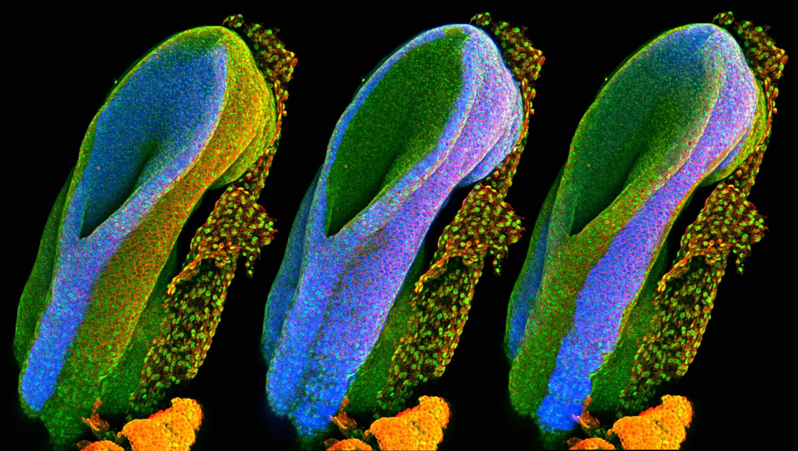
Credit: Gabriel Galea, University College London
Spinal cords are formed from a structure called the neural tube, which develops during the first month of pregnancy. These three images show the open end of a mouse’s neural tube, with each image highlighting (in blue) one of the three main embryonic tissue types. The one on the left will develop into the brain, spine and nerves; the one in the middle will form the organs; and the one on the right will eventually form the skin, teeth and hair.
Zebrafish Eye
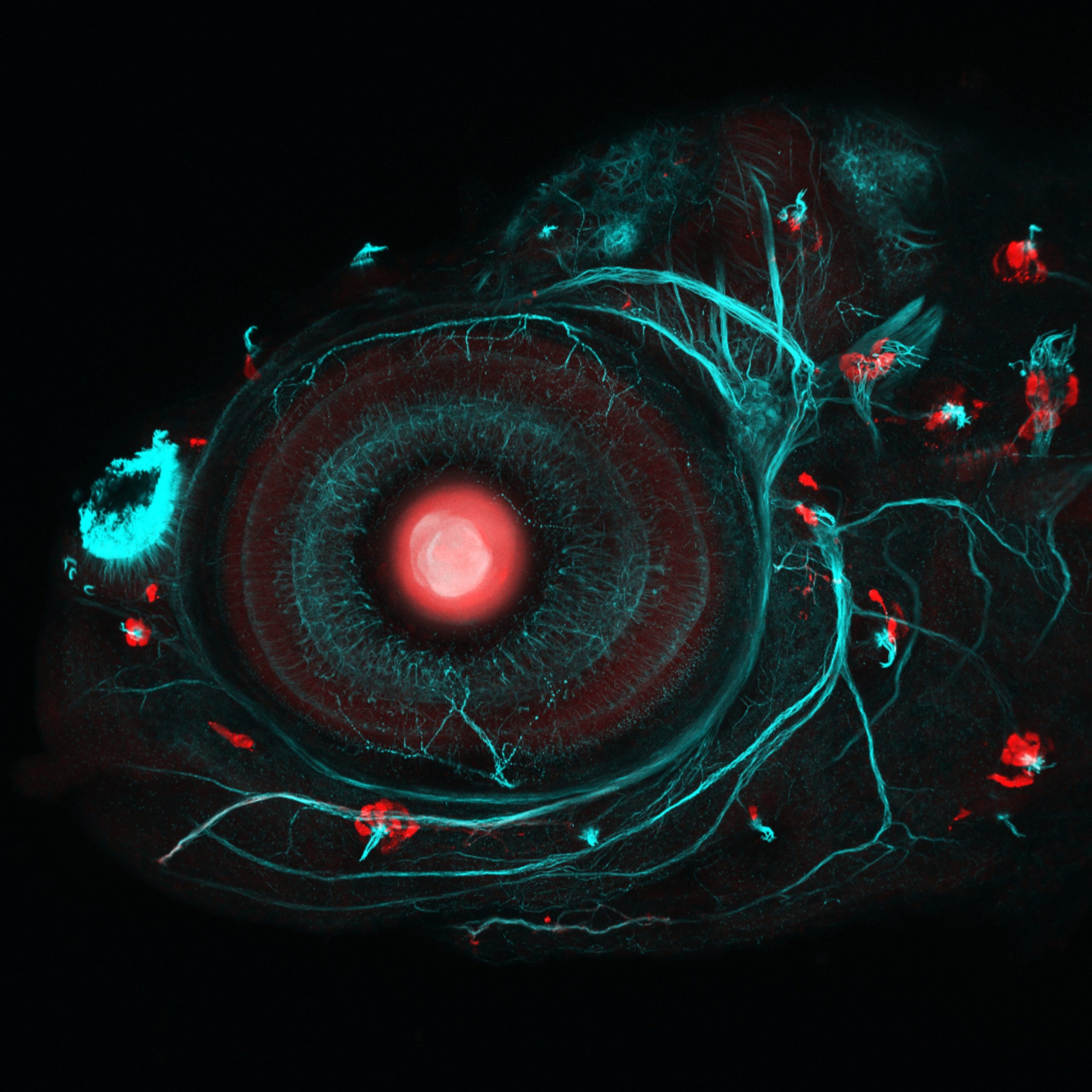
Image: Ingrid Lekk and Steve Wilson, University College London
This is the eye of a four-day-old zebrafish embryo. Using the CRISPR-Cas9 gene editing system, plus some strategic breeding, researchers from the University of College London created a zebrafish that could have specific parts of its body made to glow in fluorescent red. Here, the scientists are studying the lens of the eye (the big red dot), and cells called neuromasts (the small red dots). Neuromasts help fish respond to movements in the water, like a predator. The fish’s nervous system can be seen in greenish-blue.
Cat Skin

Credit: David Linstead
A polarised light micrograph — a type of microscopy in which filters allow only light travelling in a specific orientation to pass — of cat skin, showing hairs (yellow), whiskers (also yellow) and blood supply (black).
Iris Clip
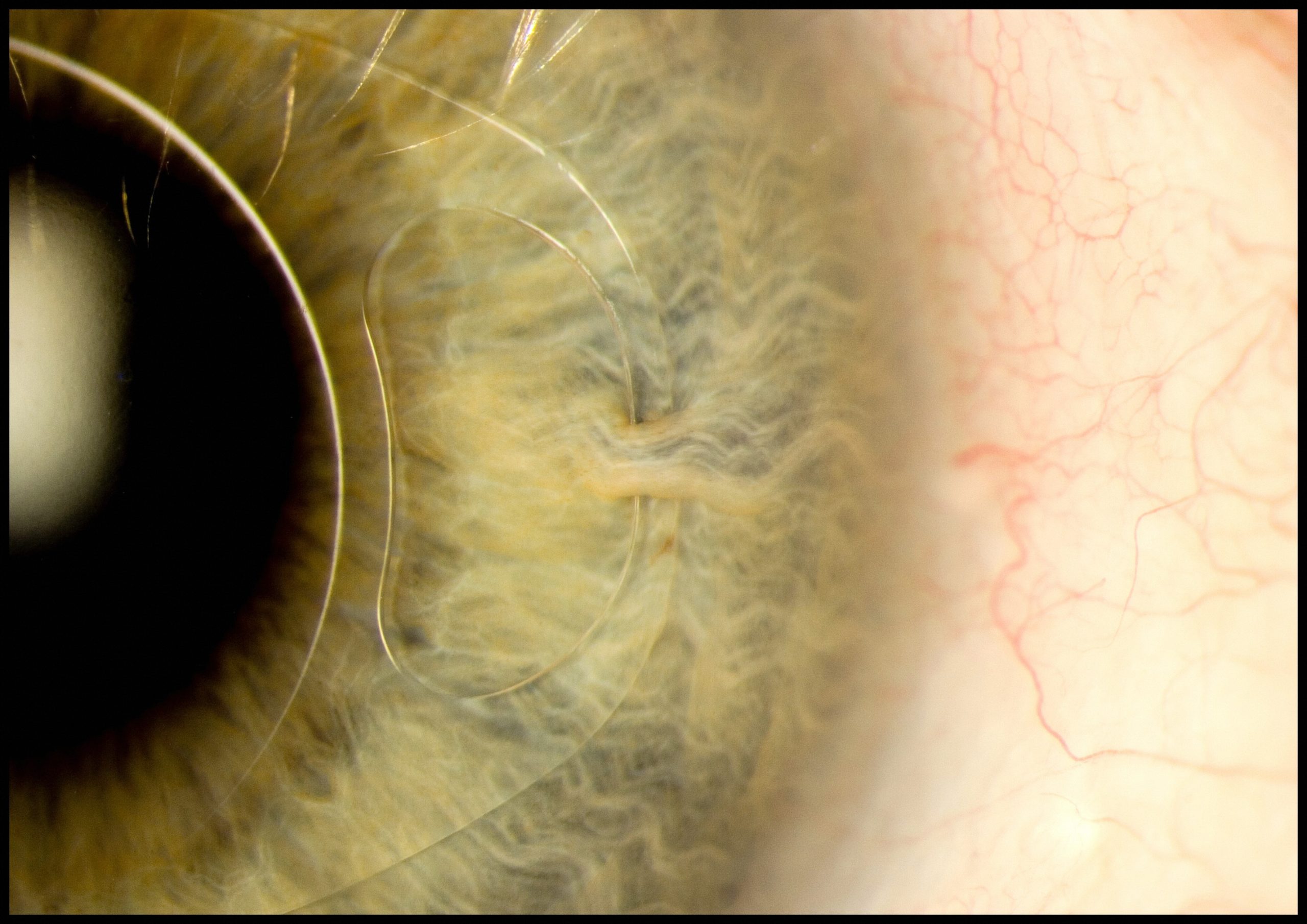
Credit: Mark Bartley, Cambridge University Hospitals NHS Foundation Trust
This image shows how an “iris clip”, also known as an artificial intraocular lens (IOL), is fitted onto the eye. The device is fixed to the iris through a small surgical incision, and is used to treat nearsightedness and cataracts.
#breastcancer Twitter Connections
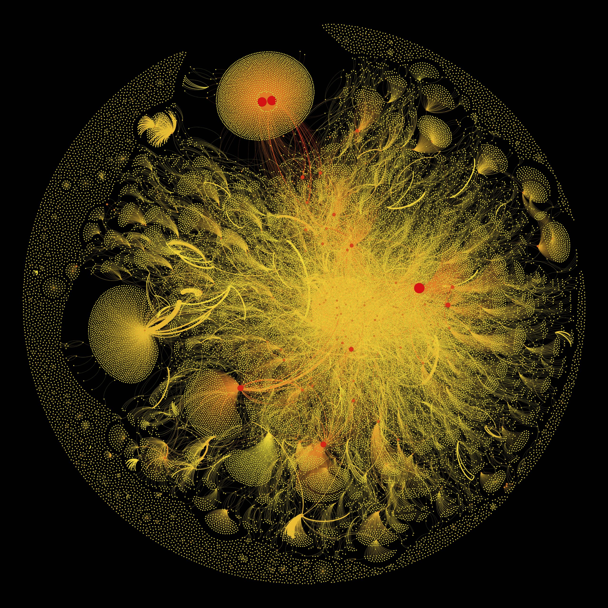
Credit: Eric Clarke, Richard Arnett and Jane Burns, Royal College of Surgeons in Ireland
A graphical visualisation of data collected from tweets containing the hashtag #breastcancer. Twitter users are represented by dots (called nodes) and lines connecting the nodes represent the relationships between the users. Nodes are sized differently according to the number and importance of the other nodes they are connected with. The thickness of each connecting line is determined by the number of times that two users interact with or mention one another. The “double yolk” structure at the top of the image (the large yellow circle containing two red dots) indicates common mentions of two accounts.
MicroRNA Scaffold Cancer Therapy
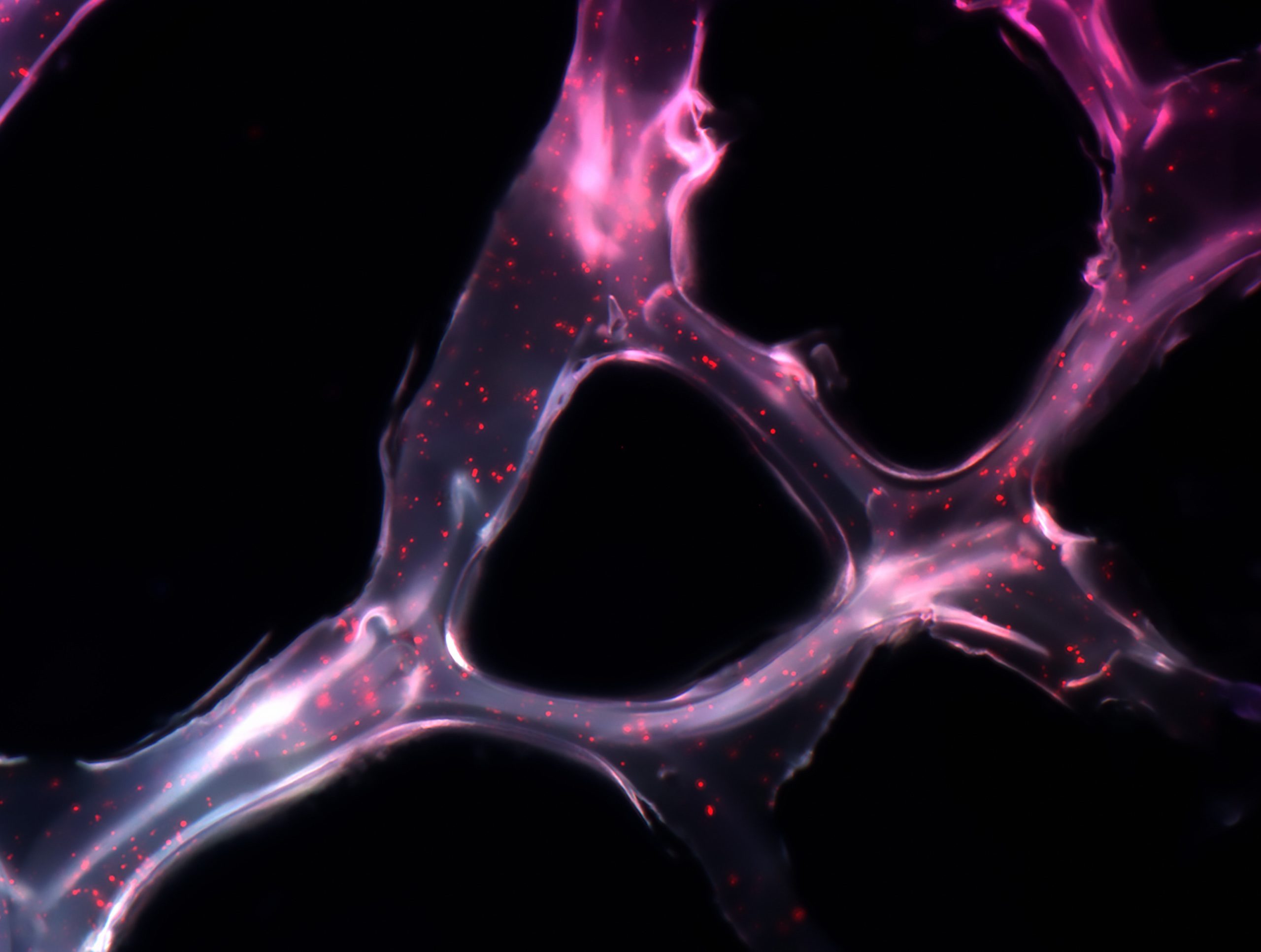
Credit: João Conde, Nuria Oliva and Natalie Artzi, Massachusetts Institute of Technology (MIT)
This synthetic net can coat a tumour and deliver short genetic sequences, called microRNAs, to cancer cells. In tests on mice, this form of cancer therapy has been shown to shrink tumours by as much as 90 per cent in just two weeks.
Hawaiian Bobtail Squid
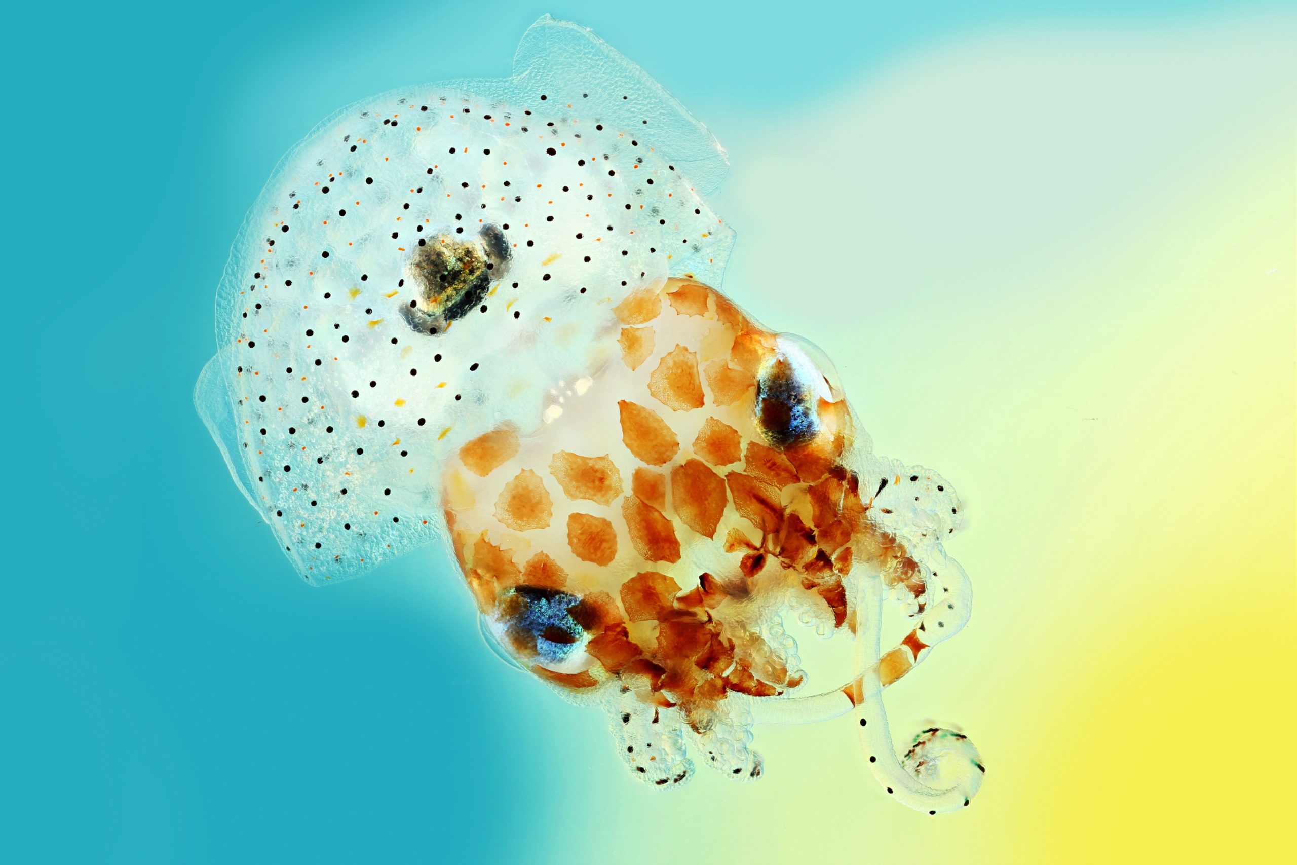
Credit: Mark R Smith, Macroscopic Solutions
Native to the Pacific Ocean, Hawaiian bobtail squid are nocturnal predators that remain buried under the sand during the day and come out to hunt for shrimp near coral reefs at night. These aquatic animals have a light organ on their underside that houses a colony of glowing bacteria called Vibrio fischeri. The squid provide food and shelter for these bacteria, in return for their bioluminescent qualities.
Pigeon Thermoregulation
Credit: Scott Echols, Scarlet Imaging and the Grey Parrot Anatomy Project
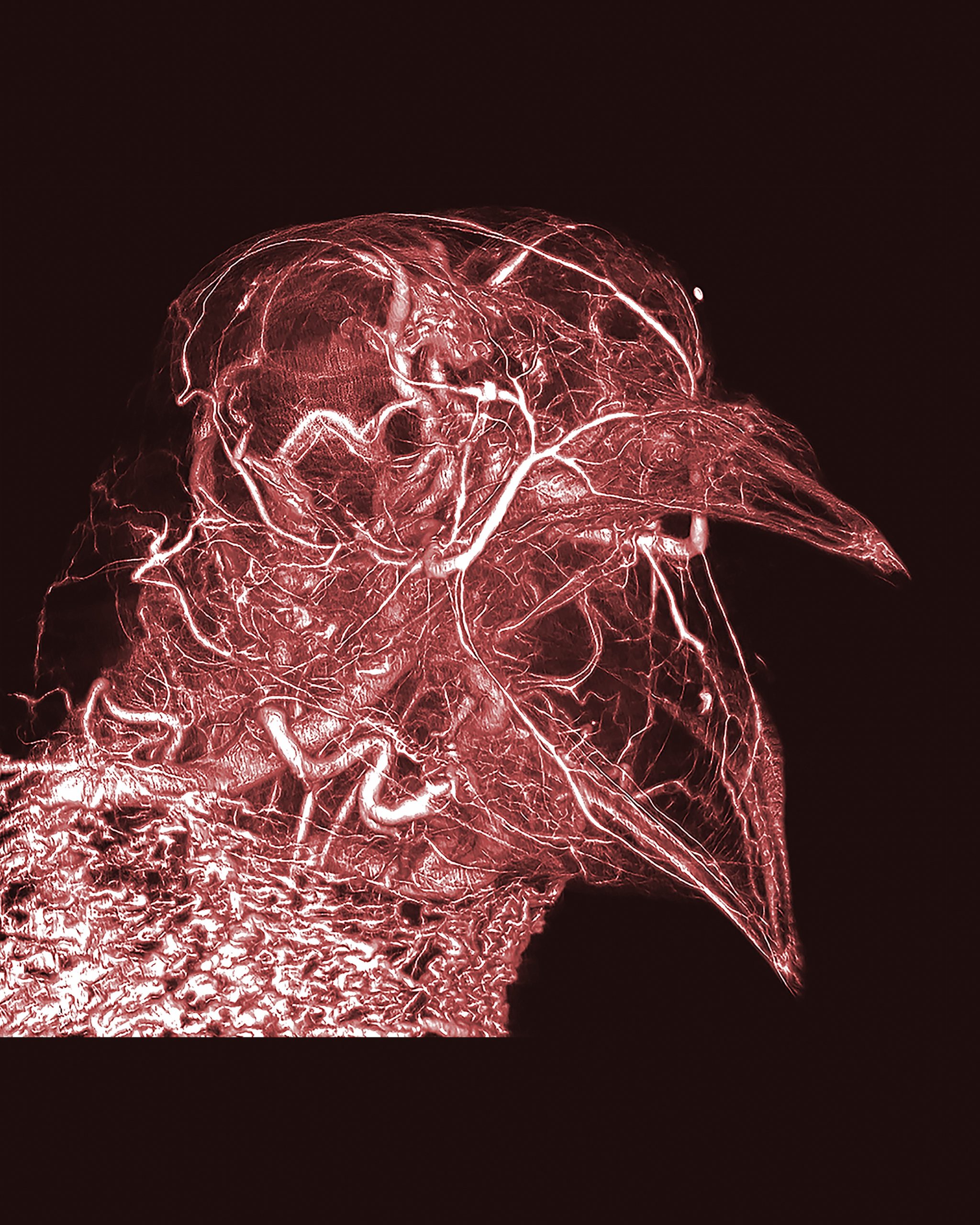
Using CT scans and digital imaging, researchers were able to see the entire network of blood vessels in a pigeon, right down to the capillary level. The intricate network of blood vessels in this pigeon’s neck can be seen at the bottom of the picture. This extensive blood supply just below the skin helps the pigeon control its body temperature through a process known as thermoregulation.
Brain on a Chip
Credit: Collin Edington and Iris Lee, © Massachusetts Institute of Technology (MIT)
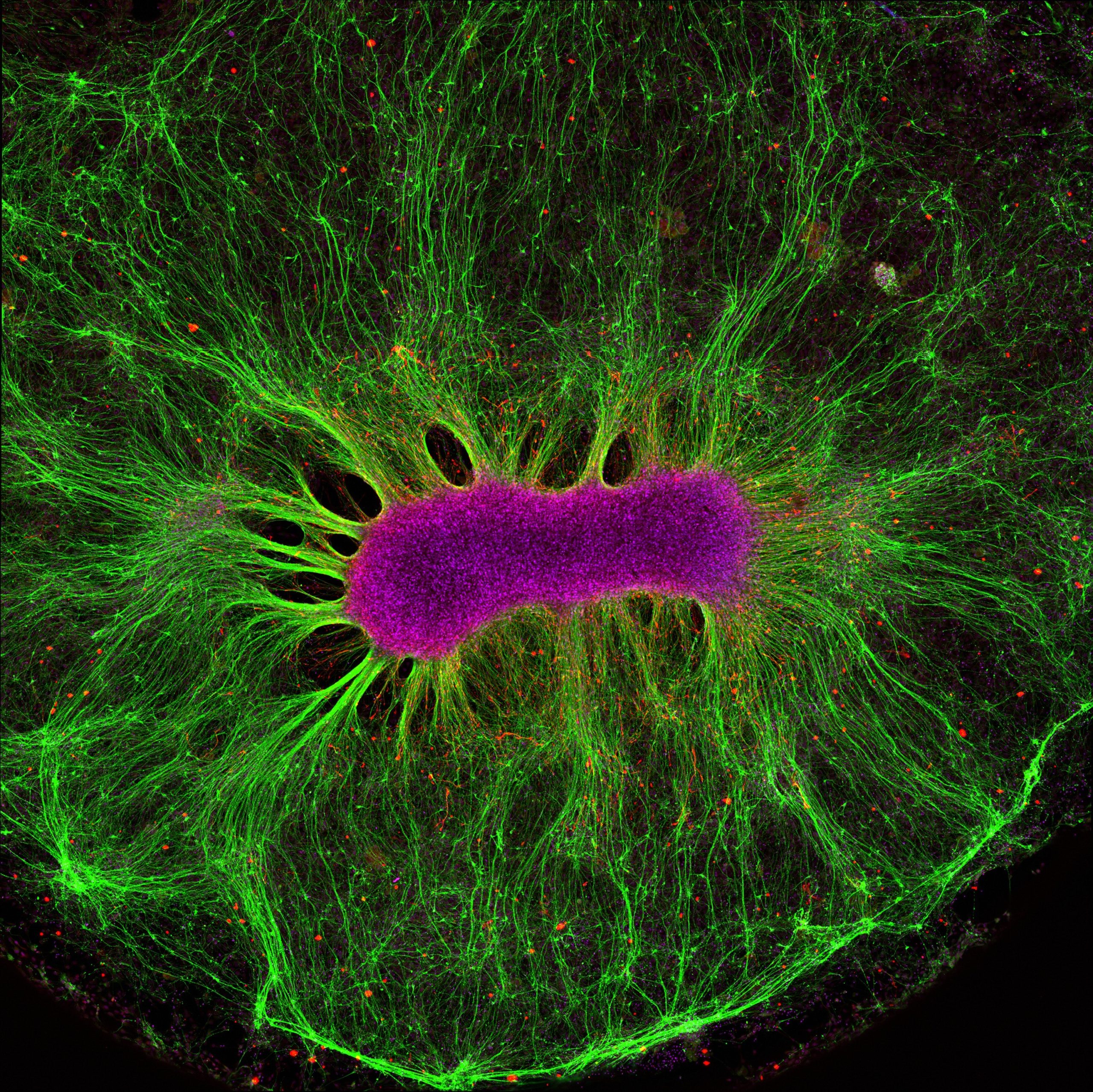
A neural stem cell grows on a synthetic gel. Though outside the body, these stem cells, shown in magenta, were able to generate nerve fibres, shown in green. Researchers are currently investigating ways of growing miniature organs on plastic chips, which could eventually become interconnected. These systems could be used to accurately predict the effectiveness and toxicity of drugs and vaccines and remove the need for animal testing in medical research.
Blood Vessels of an African Grey Parrot
Credit: Scott Birch and Scott Echols
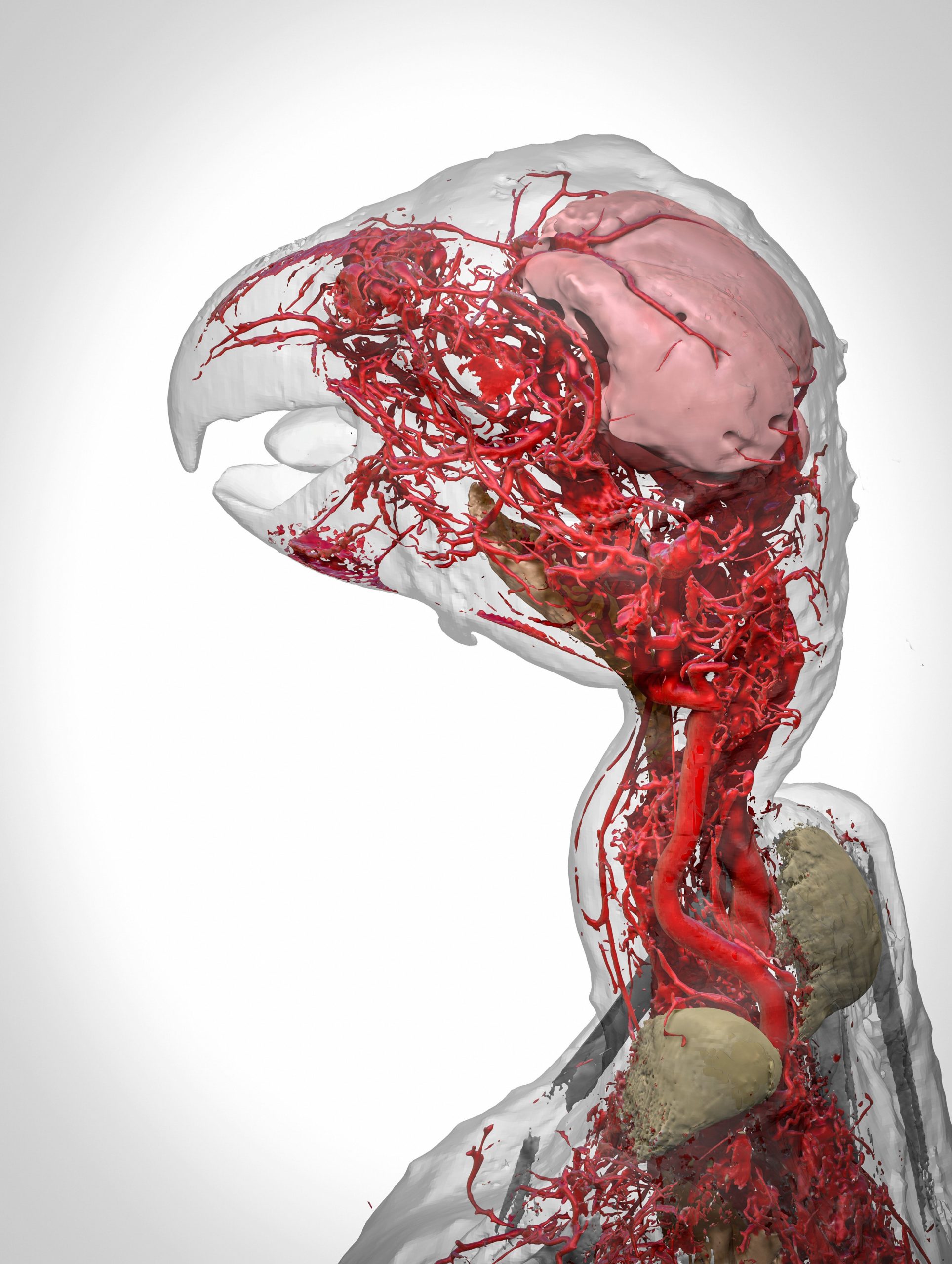
A 3D reconstruction of an African grey parrot. The model details the highly intricate system of blood vessels in the head and neck of the bird, and was made possible through the use of a new contrast agent called BriteVu. This contrast agent allows researchers to study a subject’s vascular system in incredible detail, right down to the capillary level.
The overall winner of the Wellcome Image awards will be announced on March 15.
