
Every year, the UK’s British Heart Foundation runs a competition to find the most interesting images produced by its researchers — and 2013 is a good, good year. Here are some of our favourites.
Broken Hearted
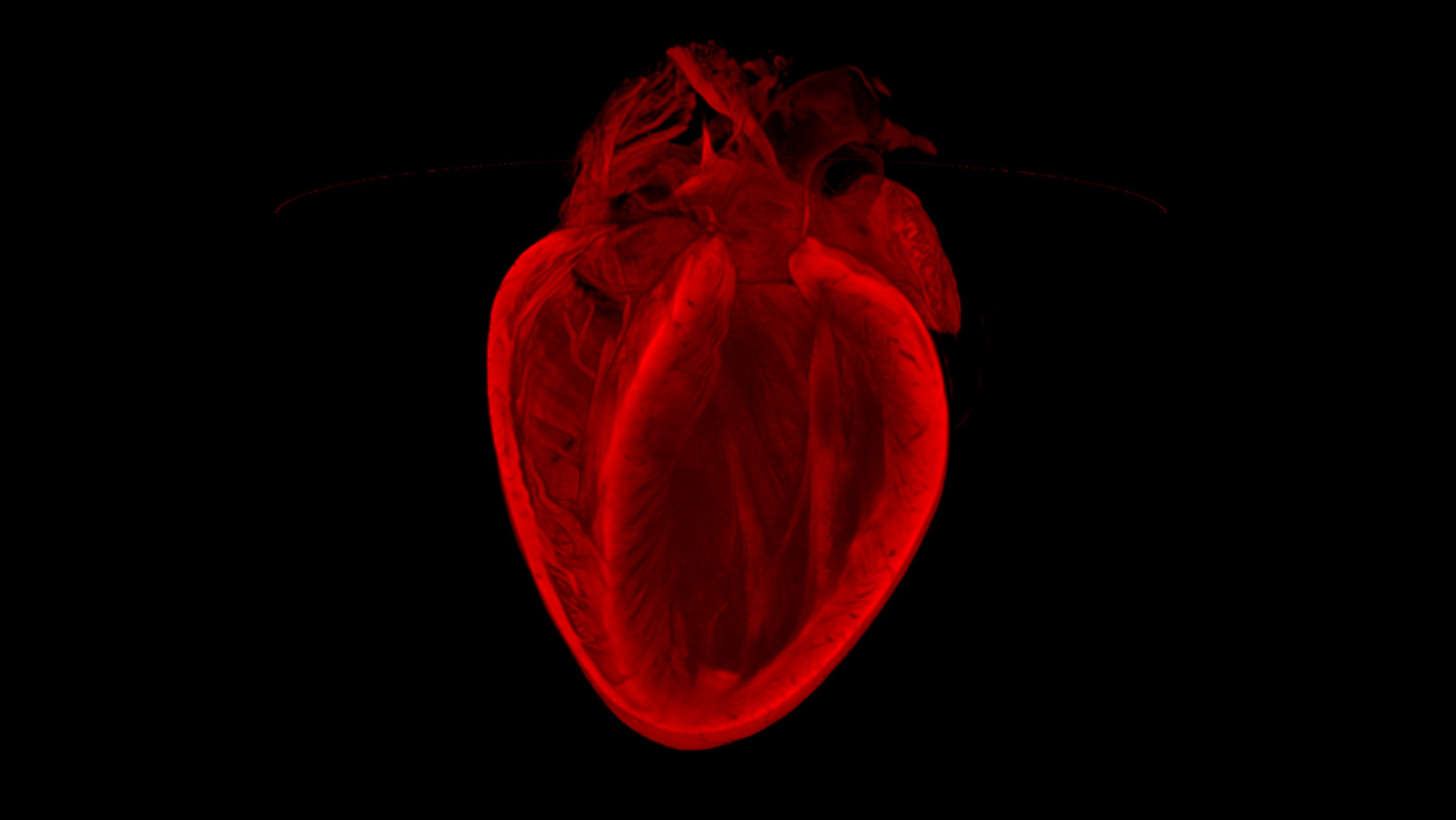
The overall winner of the BHF’s Reflections of Research competition, this image, created by Dr Gillian grey, Megan Swim and Harris Morrison form the University of Edinburgh, reveals the 3D structure of an adult mouse heart. The image was created using a technique called called Optical Projection Tomography — the optical equivalent of a CT scan. Being able to image hearts using OPT should help researchers better understand the damage caused by heart attacks.
Arrested Development
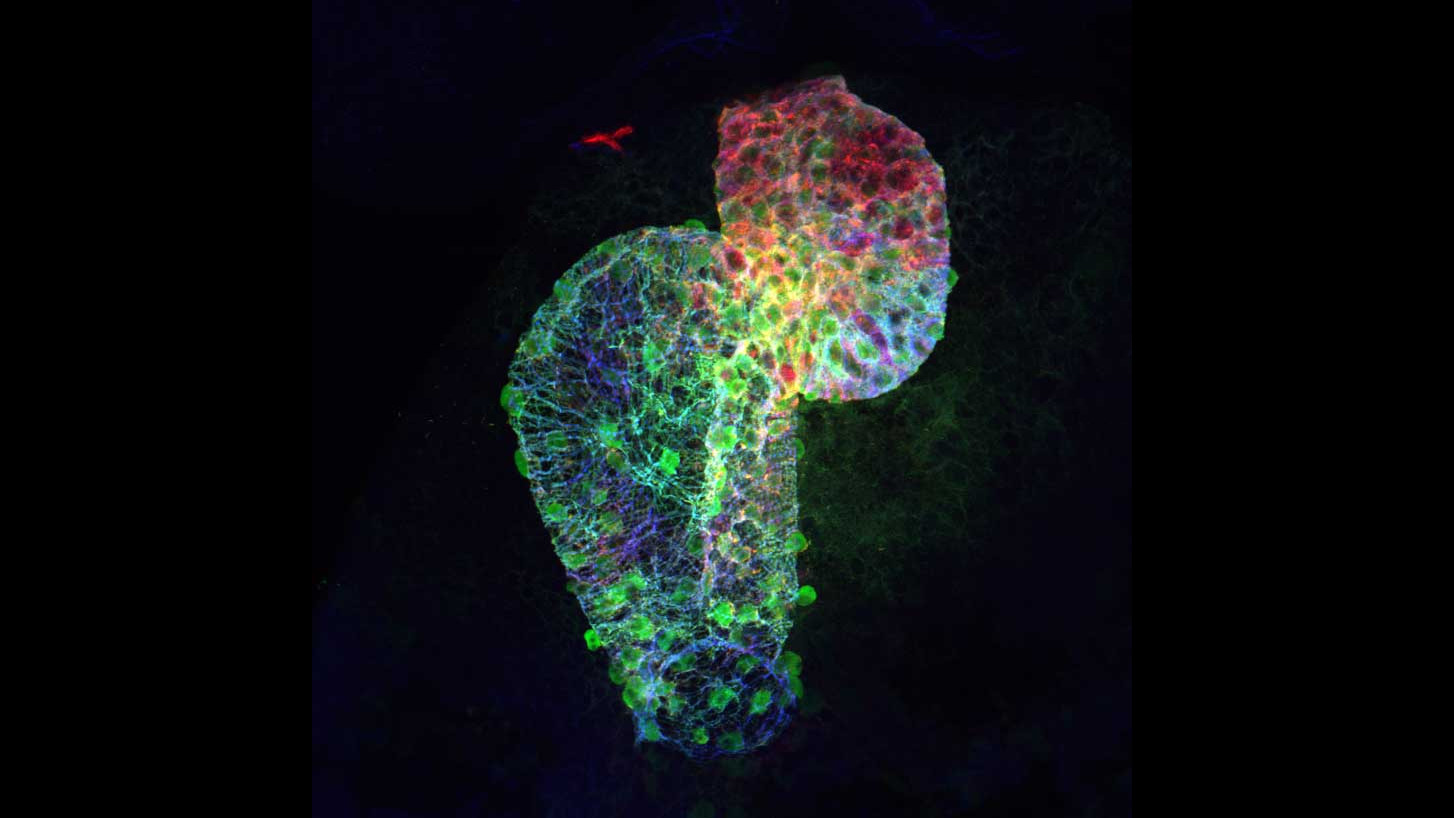
This images, acquired by Dr Jana Koth from the University of Oxford, may look like a wild LSD trip, but it is in fact the front view of a developing two-day-old zebrafish heart. The green patches are heart muscle cells, while the blue and red spots are material will go on to form said cells. You can make out two sections that have already formed: a large, thin atrium (where blood flows in) and small, thick ventricle (where blood flows out).
True Romance
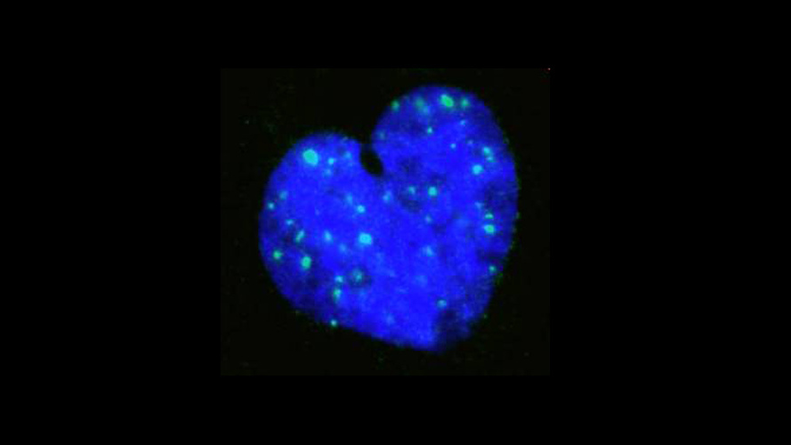
From actual hearts to cells that look like hearts: this romantic image, captured by Dr Andrew Cobb, King’s College London, is of the heart-shaped nucleus of a single vascular smooth muscle cell. In case you’re wondering, they’re the cells that give blood vessels their shape. This particular nucleus’ shape is unusual — which could be explained by the green specks, which indicate regions of DNA damage.
Killer Cholesterol
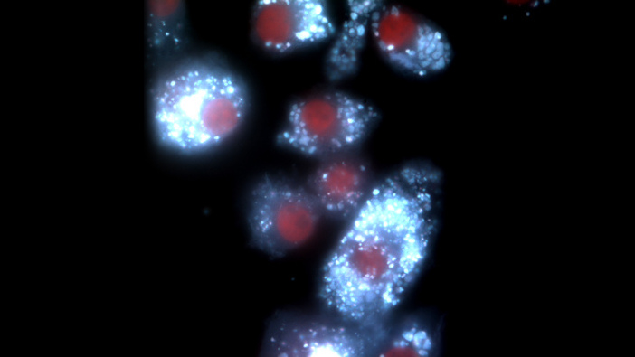
Looking more like something from outer space than inner body, this image, by Dr Yichuan Wen and Dr David Leake from the University of Reading, actually shows a special type of immune cells called foam cells. Present in the arteries of people with atherosclerosis, those innocent-looking white specks are actually cholesterol, which will go on to be altered by the cells — and possibly lead to heart attacks and strokes.
Pictures: British Heart Foundation