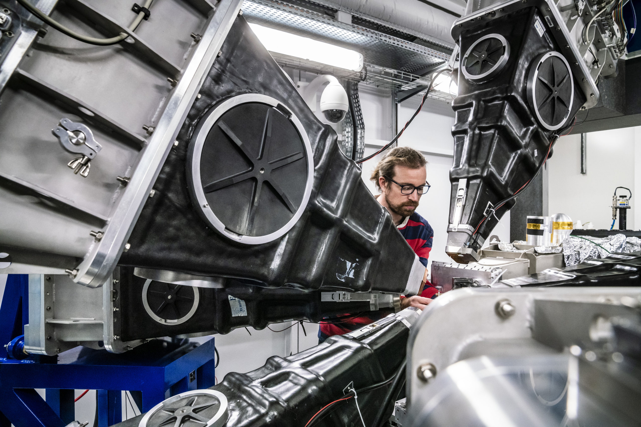
A new way of producing powerful X-ray beams — the brightest on Earth — is now making it possible to create 3D images of matter at astounding resolutions. This “Extremely Brilliant Source” officially opened last month at the European Synchrotron Radiation Facility in France, and scientists are already using it to study the coronavirus behind covid-19. These X-ray beams will image the interiors of fossils, brains, batteries, and countless other interesting items down to the atomic scale, revealing unprecedented information and supercharging scientific research.
A typical medical X-ray, like you would get for a broken bone, can show doctors details about your particular fracture and the tissue around it. X-rays penetrate the body and are absorbed at different rates by different tissue; once they’ve passed through you, they hit a detector, creating the familiar black-and-white X-ray image. The Extremely Brilliant Source produces X-rays 10 trillion times more powerful than those used in hospitals. With such a beam, scientists could create a 3D image of your broken bone so detailed that they could see the individual atoms in the blood cells surrounding your fracture. Of course, you wouldn’t want to be hit with this particular beam — the dose of radiation would be fatal.
The possibilities that the Extremely Brilliant Source opens up feel endless. One area that particularly excites Francesco Sette, director general of the ESRF, is research into the structure and functioning of brains, which could eventually enable brain-like electronics. “It would be a major revolution, not only for neuroscience, but also for all those applications that are coming up to use possibly the human brain architecture for a new generation of devices,” he said.
Using synchrotron X-ray imaging, engineers can gain minute insights into innovative materials, aiding fields like aeronautics and nanoelectronics. Paleontologists can study the tiny interior structures of fossils without needing to destroy their samples. This summer, some of the first researchers to have access to the Extremely Brilliant Source used it to image the complete lungs of people who died of covid-19, and they were able to identify previously unseen damage caused by the virus at the microscopic level.
A synchrotron is simply a particle accelerator that uses magnetic fields to accelerate charged electrons to such high energies that they emit X-ray light, also known as synchrotron light. (Unlike at the Large Hadron Collider, for example, the particles zipping around a synchrotron loop aren’t made to collide with each other.) The X-rays produced by the rapidly circulating electrons are siphoned out of the accelerator ring and into 44 specialised laboratories, known as beamlines. Researchers then use those beams to image their targets. In recent decades, synchrotron-based science has been behind all manner of breakthroughs, including recently allowing researchers to see inside an unhatched dinosaur egg and to read an ancient book destroyed by a volcano.
The European Synchrotron Radiation Facility in Grenoble, France, has been operating since 1994. The previous iteration of its X-ray source was already the most powerful in the world; this year’s upgrade increases its power by a factor of 100. The facility shut down in December 2018 to begin the transition to the Extremely Brilliant Source. Fortunately, the covid-19 pandemic didn’t delay the official opening on August 25, as the project was running nearly five months ahead of schedule. Researchers are already using the beams, and the very first published results from recent work at the synchrotron should be out soon, according to Sette.
What made this significant upgrade possible was a new design for a lattice of 1,100 magnets that drive the electrons around the 844-metre-round ring. These magnets not only accelerate the electrons forward but also give them slight “kicks,” changing their direction. Those small changes in direction are the key to producing the X-rays.

“When you deviate the trajectory of a charged particle, you produce light,” Sette explained. “And this light is what we call synchrotron light.”
It’s a simple matter of the law of conservation of energy: When you bend an electron beam so that it travels in a loop instead of a straight line, the electrons lose a bit of energy every time they change direction. That lost energy is in the form of light. To get the emitted light to be in the X-ray frequency range, you need to provide a stronger magnetic “kick.” The new magnet lattice design makes it possible to continuously bend and refocus the electron beam, producing large amounts of high-energy X-ray light without requiring a bigger ring facility.
A field that could be dramatically pushed forward by synchrotron science is histology, the microscopic study of tissues. Today, histologists study tissues by slicing them into many extremely thin samples and staining them with dyes to reveal microscopic structures. With synchrotron imaging, samples don’t need to be sliced and stained; researchers can image them whole, creating high-resolution, 3D scans that show so much more about their anatomy.
“This has been dubbed ‘3D nano-histology,’ which is a dream for the medical world,” Sette said. “It represents a complete revolution in performing histology.”
One scientist who has done research using the previous iteration of the European Synchrotron is Victor Gonzalez, a postdoctoral researcher at the Rijksmuseum Science Department in Amsterdam. Gonzalez regularly uses synchrotron imaging to study centuries-old paint samples. His work recently uncovered details about the painting techniques of Rembrandt.
“For my research community, the ESRF upgrade is very important,” Gonzalez said in an email. “The new state-of-the-art analytical capacities of the facility will allow us to analyse precious samples faster than ever. An experiment that took several days before will now be conducted in just an afternoon! For us it means a wealth of data suddenly available, so new opportunities to understand the chemical mechanisms at stake in historical paint layers.”
Now that the Extremely Brilliant Source is up and running, scientists around the world can apply for time on the beamlines. It’s a competitive process — a research application must go through peer review before scientists gain access to the synchrotron. But with the new upgrade, experiments that would have taken weeks can now be done in a day; what once took a day will take just minutes. Stay tuned for plenty of exciting new science in the coming months.
