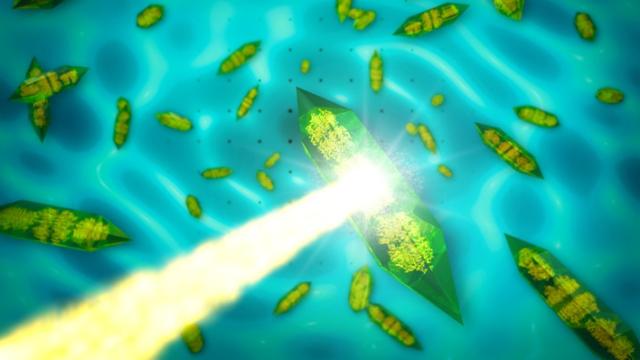
There is now a better way to image the internal structure of biological molecules at the atomic scale, using powerful X-ray lasers. This could eventually lead to important new innovations in clean energy technologies and drug development, among other uses.
Crystals are defined by their regular, lattice-like arrays of atoms, and scientists typically rely on a technique called X-ray crystallography to image that internal structure. But the more disordered the crystal, the poorer the resolution of the image. Physicists at DESY in Germany and the Linac Coherent Light Source (LCLS) facility at SLAC National Accelerator Laboratory have combined crystallography with single-molecule imaging to take very high resolution images even with disordered crystals. Their results were published yesterday in Nature.
The field of X-ray crystallography dates back to the early 20th century. Since then, scientists have used this method to figure out the atomic structure of a vast number of materials in physics, chemistry and biology. Shine X-rays onto a crystal and they will bounce off the atoms that make up the crystal. This scattering can be picked up by a detector, and the resulting diffraction pattern will have a lot of bright spots. These are known as “Bragg peaks”. By measuring those peaks, scientists can infer the crystal’s internal structure.
Sebastien Boutet, an equipment scientist at LCLS and co-author on the paper, draws an analogy to a re-planted forest with trees lined up in identical perfect rows. “Crystallography would be like standing in front of that forest and kicking billions of soccer balls at it,” he told Gizmodo via email. Some soccer balls will hit the first tree in a row, while others will penetrate deeper into the forest and hit other trees. “After they hit a tree, there is only a certain set of directions they can bounce and still find a way out of the forest, without hitting another tree,” said Boutet.
Those angles of direction are akin to Bragg peaks. Scientists can count up how many “soccer balls” emerged from each direction to determine the size and shape of the tree. Granted, it’s an imperfect analogy; there’s probably an easier way to measure the trees in a forest. But for tiny objects like atoms in a biomolecule like a protein, scientists must resort to different methods, and crystallography has proved its usefulness time and time again over the last century.
If you had a perfectly ordered crystal, you’d get nothing but Bragg’s peaks. But it’s really hard to get all those metaphorical trees lined up so precisely. “When the crystals are disordered, the Bragg peaks can no longer exist, because the array of trees is not perfect,” said Boutet.
Artistic representation of an imperfect crystal, with repeating units portrayed as a series of infinite ducks. Credit: SLAC National Accelerator Laboratory.
Even though you don’t get Bragg peaks with disordered crystals, all that X-ray energy you’ve zapped them with still bounces back out, except this time it forms a pattern called continuous diffraction.
To revisit the forest analogy, “Instead of kicking the ball at an entire forest to figure out the average shape of a tree, it would be a lot easier to kick the ball at a single tree, and figure out the shape from where the balls bounce off,” said Boutet, much like what happens with single molecular imaging. “In this case, because the balls bouncing off the single tree would not be limited to a few angles, they would not collect at single points (Bragg peaks) but would instead be more evenly distributed (continuous diffraction).”
It turns out that this latter pattern contains much more information about a molecule’s structure, because there is information not just at the Bragg peaks, but in between them as well. This in turn yields better resolution than has been possible using traditional crystallography. In essence, imperfect (disordered) crystals have both Bragg peaks and the non-Bragg signals of single molecular imaging, so this new approach exploits the best of both worlds.
Comparison of image resolution achieved by conventional crystallography (left) and the combination method (right). Credit: Dominik Oberthuer and Kartik Ayyer, DESY.
Knowing the precise structure of biological molecules is critical when it comes to real-world applications. Take drug design and development as a case in point. The goal here is to design molecules to counteract those that give rise to diseases. Boutet compares it to a lock and key. A virus, for example, has a “hole” or “lock” by which it can attach itself to a corresponding “key” on certain vital molecules in your body. This effectively disables them and allows the virus to spread. If you could design a drug whose molecules effectively plug that hole, the virus would be disabled instead. But to do that, you’d need to know the precise locations of all the atoms in those molecules.
So far the team has used their method on just one kind of biomolecule: photosystem II. They chose it because it is highly efficient at photosynthesis: using sunlight to turn water into oxygen while providing energy for plants or bacteria. “No man-made system approaches the efficiency level achieved by nature in this case,” said Boutet. “So this could be a path to developing new [clean] energy technologies, by copying nature.”
They are confident they can extend their method to many other kinds of biomolecules in the future. One potential stumbling block is whether this technique will work at other facilities. X-ray lasers like the one at LCLS are extremely powerful, enabling the researchers to take data very quickly. This is important because the longer the sample must be exposed to the X-rays, the higher the risk of damage. Other X-ray sources, like synchrotrons, aren’t as powerful so the samples would need longer exposures.
However, “Once the full potential of the new method is understood, it could turn out to be one of the biggest advances since the birth of crystallography,” LCLS director Mike Dunne said in a statement.
[Nature]
Top image: Artist’s representation of X-rays passing through a crystal. Credit: SLAC National Accelerator Laboratory.
