Researchers from the University of Washington are the first to visualise the insidious way that the flu virus latches onto a cell and plows its way inside, causing an infection.
Image: Kelly Lee Lab
Using advanced electron microscopy, a team led by Kelly Lee sequenced the stages of membrane fusion — the process in which two separate biological entities merge to become one. Specifically, they chronicled the merger of the influenza virus and a small lipid vesicle (or liposome), which was used as a stand-in for a cellular membrane. (Using a whole cell was impractical for the type of microscopy used in the study.) Their results now appear in the latest edition of the Journal of Virology.
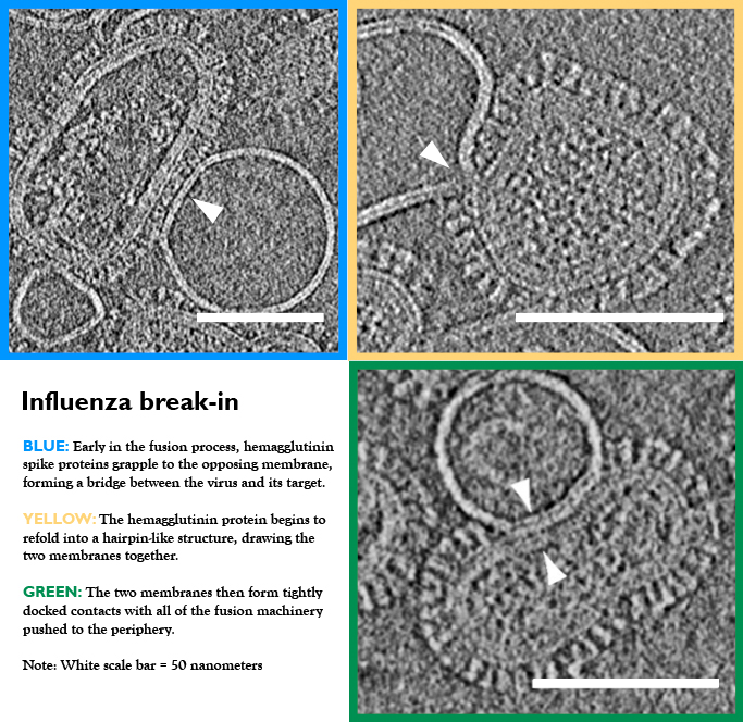
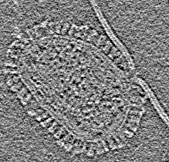
Image: Kelly Lee Lab
“What we’ve done is to image the different steps that take place as the virus’ fusion machinery start to manipulate the target membrane barrier so as to be able to open up a hole and spill its genome cargo into the cell,” Lee told Gizmodo.
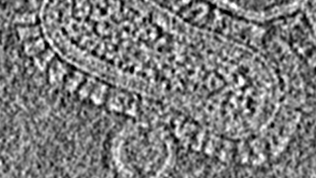
This process occurs over the course of several stages. First, the flu virus throws out short peptide segments that essentially work like grappling hooks. These peptides grab onto the target membrane, and then alter their structure to pinch together the two membranes.
The virus then snuggles up against its target, creating even larger zones of contact between the two objects. These contact sites begin to unzip and break down, opening up a channel from the virus into the interior of the liposome. The virus then delivers its genome cargo to start a new infection.
A gap opens up between the virus and the membrane. (Image: Kelly Lee Lab)
“Basically, these viruses have evolved nanoscopic machines that respond to triggers and change their structure like transformers,” said Lee. “They really are tiny machines that carry out surgery at the nanoscopic biological scale.”
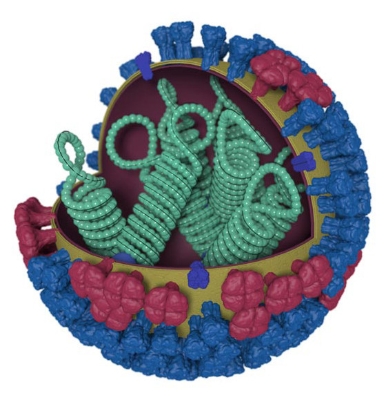
Conceptual image of a flu virus. The outer layer of blue spikes latch on to target cells. (Image: CDC)
The researchers say that the exact same process happens when the virus attacks and infiltrates a real, full-sized cell. Any virus that has a membrane — such as the flu, herpes, HIV, measles and ebola — is equipped with similar molecular machinery. This enables them to invade cells — and they do so by ripping open the membrane barriers that are in its way.
Antibodies can target this machinery, locking them into place so that the virus can’t change its structure or preventing it from binding to the target.
In addition to helping with research into these antibodies, Lee hopes that his team’s work will also help to improve the understanding of other critical biological functions, such as sperm-egg fertilisation and signalling across nerve synapses.